. 2017 Aug 10:259:128-135.
doi: 10.1016/j.jconrel.2017.03.006. Epub 2017 Mar 6.

PBCA-based polymeric microbubbles for molecular imaging and drug delivery

Affiliations

PBCA-based polymeric microbubbles for molecular imaging and drug delivery

Patrick Koczera et al. J Control Release. .

Abstract

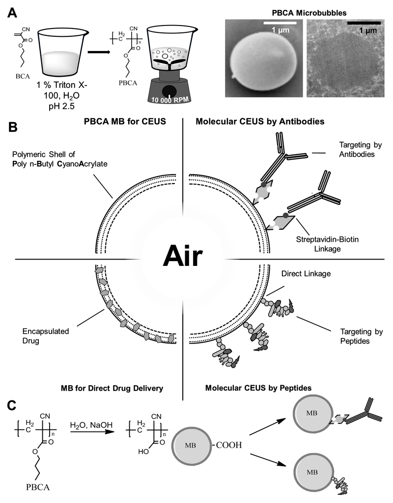
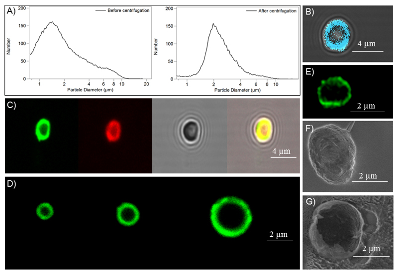
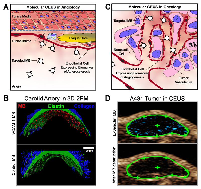
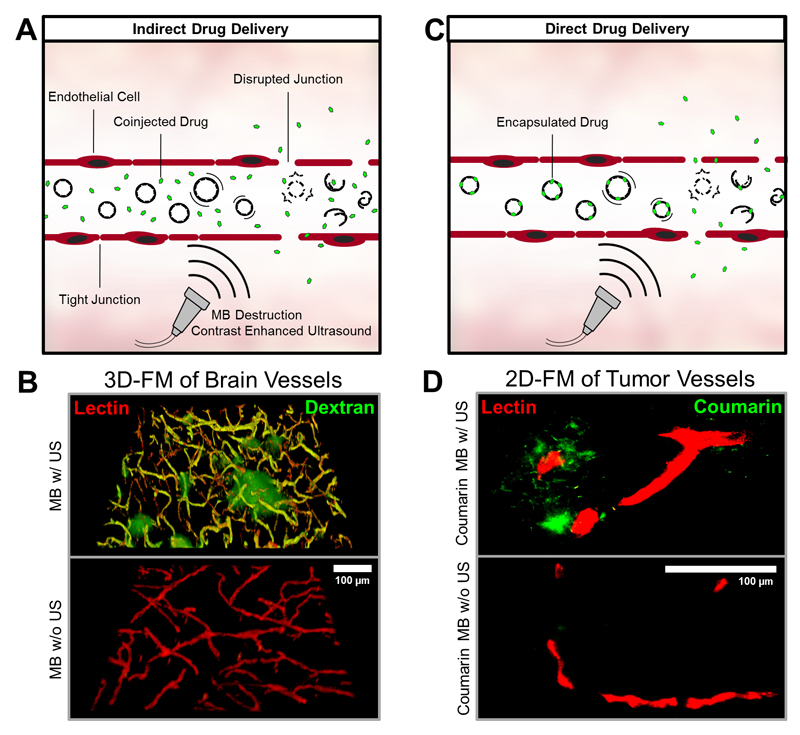
Microbubbles (MB) are routinely used as contrast agents for ultrasound (US) imaging. We describe different types of targeted and drug-loaded poly(n-butyl cyanoacrylate) (PBCA) MB, and demonstrate their suitability for multiple biomedical applications, including molecular US imaging and US-mediated drug delivery. Molecular imaging of angiogenic tumor blood vessels and inflamed atherosclerotic endothelium is performed by modifying the surface of PBCA MB with peptides and antibodies recognizing E-selectin and VCAM-1. Stable and inertial cavitation of PBCA MB enables sonoporation and permeabilization of blood vessels in tumors and in the brain, which can be employed for direct and indirect drug delivery. Direct drug delivery is based on US-induced release of (model) drug molecules from the MB shell. Indirect drug delivery refers to US- and MB-mediated enhancement of extravasation and penetration of co-administered drugs and drug delivery systems. These findings are in line with recently reported pioneering proof-of-principle studies showing the usefulness of (phospholipid) MB for molecular US imaging and sonoporation-enhanced drug delivery in patients. They aim to exemplify the potential and the broad applicability of combining MB with US to improve disease diagnosis and therapy.

Keywords: Microbubbles; Nanomedicine; Sonoporation; Tumor targeting; Ultrasound.


Figures

Figure 1
Figure 1. Synthesis and functionalization of PBCA-based polymeric MB.
A: BCA monomers are added to an aqueous solution containing the surfactant Triton X-100 at pH 2.5. Vigorous stirring creates polydisperse MB with PBCA as the shell material. Scanning (SEM) and transmission (TEM) electron microscopy exemplify the spherical shape of PBCA MB. B: Schematic depiction of the surface-functionalization and shell-loading of PBCA MB, making them useful for molecular imaging and drug delivery. Coupling of antibodies and peptides as targeting moieties for molecular imaging targeted drug delivery can be performed by biotin-streptavidin linkages, as well as via direct chemical coupling mechanisms. C: For surface-functionalization, the shell of PBCA MB is hydrolyzed to create carboxyl-groups for chemical coupling to targeting ligands.
Figure 2
Figure 2. Characterization and loading of PBCA-based polymeric MB.
A: Size distribution of MB before and after centrifugation and flotation. B-D: Fluorescence microscopy images of HITC-loaded MB (B), individual and co-loading of rhodamine B and coumarin 6 into MB (C), and differently-sized MB batches loaded with coumarin 6 (D). E: STED microscopy image of a coumarin 6-loaded MB. F-G: Scanning electron microscopy (SEM) images of an intact (F) and a destroyed (G) PBCA MB.
Figure 3
Figure 3. PBCA-based polymeric MB for molecular imaging.
A: Schematic depiction of the retention of ligand-targeted MB for contrast-enhanced US imaging of endothelial biomarkers in atherosclerosis. B: Ex vivo binding of rhodamine-labeled VCAM-1-targeted MB to an explanted murine carotid artery after TNF-α stimulation, as assessed by 3D two-photon laser scanning microscopy (3D-2PM). VCAM-1-targeted MB bind to activated endothelial cells and show retention under physiological shear stress (upper panel), whereas untargeted MB hardly associate with inflamed blood vessels (lower panel). Rhodamine-labeled MB are shown in red, elastin autofluorescence in green, and second harmonic generation imaging of collagen is depicted in blue. C: Illustration of the binding of targeted MB for contrast-enhanced US of angiogenesis biomarkers during tumor progression. D: Non-linear molecular US imaging of peptide-modified MB recognizing E-selectin on tumor blood vessels in subcutaneous A431 xenograft tumors. Signal enhancement (in blue) as a difference of contrast intensity before (upper panel) and after (lower panel) MB destruction by destructive US pulses allows for the assessment of E-selectin expression, and analysis of tumor vascularization and angiogenesis.
Figure 4
Figure 4. PBCA-based polymeric MB for indirect and direct drug delivery.
A+C: After i.v. injection, MB can be used to enhance the permeability and penetration of co-administered (A: indirect drug delivery) or co-formulated (B: direct drug delivery) drugs and drug delivery systems. B: Ex vivo two-photon laser scanning microscopy (3D-FM) of the extravasation of the macromolecular model drug FITC-dextran (green) across the blood-brain barrier. Rhodamine-lectin-stained blood vessels are shown red. Upon US-induced MB destruction (upper panel), the accumulation and penetration of FITC-dextran can be clearly detected in the mouse brain. No FITC-dextran extravasation is observed if US is omitted (lower panel). D: Ex vivo fluorescence microscopy (2D-FM) images of coumarin 6 (green) accumulation, released from the MB shell or entrapped within MB shell fragments, in subcutaneous CT26 colon carcinoma tumors in mice, in relation to rhodamine-lectin-stained tumor blood vessels (red). Upon US-mediated MB destruction of VEGFR-2-targeted and coumarin 6-loaded MB, substantially enhanced model drug delivery to and into tumorous tissue can be observed.

References

    1. Wells PNT. Ultrasound imaging. Phys Med Biol. 2006;51(13):83. - PubMed
    1. Bauer A, et al. Wideband harmonic imaging: A novel contrast ultrasound imaging technique. Eur Radiol. 1999;9(3):364–367. - PubMed
    1. Tiemann K, et al. Stimulated acoustic emission: pseudo-Doppler shifts seen during the destruction of nonmoving microbubbles. Ultrasound in Medicine & Biology. 2000;26( 7):1161–1167. - PubMed
    1. Cosgrove D. Future Prospects for SonoVue and CPS. Eur Radiol. 2004;14(Suppl 8):116–124. - PubMed
    1. Ehling J, Lammers T, Kiessling F. Non-Invasive Imaging for Studying Anti-Angiogenic Therapy Effects. Thromb Haemost. 2013;109(3):375–390. - PMC - PubMed

Publication types

MeSH terms