Comparative Study
. 2017 Apr;38(4):729-734.
doi: 10.3174/ajnr.A5137. Epub 2017 Mar 9.

Comparison of the Diagnostic Utility of 4D-DSA with Conventional 2D- and 3D-DSA in the Diagnosis of Cerebrovascular Abnormalities

Affiliations
Comparative Study

Comparison of the Diagnostic Utility of 4D-DSA with Conventional 2D- and 3D-DSA in the Diagnosis of Cerebrovascular Abnormalities

C Sandoval-Garcia et al. AJNR Am J Neuroradiol. 2017 Apr.

Abstract

Background and purpose: 4D-DSA is a time-resolved technique that allows viewing of a contrast bolus at any time and from any desired viewing angle. Our hypothesis was that the information content in a 4D-DSA reconstruction was essentially equivalent to that in a combination of 2D acquisitions and a 3D-DSA reconstruction.

Materials and methods: Twenty-six consecutive patients who had both 2D- and 3D-DSA acquisitions were included in the study. The angiography report was used to obtain diagnoses and characteristics of abnormalities. Diagnoses included AVM/AVFs, aneurysms, stenosis, and healthy individuals. 4D-DSA reconstructions were independently reviewed by 3 experienced observers who had no part in the clinical care. Using an electronic evaluation form, these observers recorded their assessments based only on the 4D reconstructions. The clinical evaluations were then compared with the 4D evaluations for diagnosis and lesion characteristics.

Results: Results showed both interrater and interclass agreements (κ = 0.813 and 0.858). Comparing the 4D diagnosis with the clinical diagnosis for the 3 observers yielded κ values of 0.906, 0.912, and 0.906. The κ values for agreement among the 3 observers for the type of abnormality were 0.949, 0.845, and 0.895. There was complete agreement on the presence of an abnormality between the clinical and 4D-DSA in 23/26 cases. In 2 cases, there were conflicting opinions.

Conclusions: In this study, the information content of 4D-DSA reconstructions was largely equivalent to that of the combined 2D/3D studies. The availability of 4D-DSA should reduce the requirement for 2D-DSA acquisitions.


Figures

Fig 1.
Fig 1.
Screenshot of the evaluation form used in the evaluation of the 4D-DSA reconstructions.
Fig 2.
Fig 2.
A comparison of conventional 3D-DSA (upper row) with different timeframes from a 4D-DSA reconstruction (lower row). The projections of the 2 image types are identical. The 3D-DSA images allow viewing from any desired angle; however, in this case because of the vascular overlap, it is impossible to clearly see the relationship of the small aneurysm (yellow circle) to its parent artery. Because the 4D images allow viewing not only from any desired angle but also at any time during the passage of a contrast bolus through the vasculature, the early timeframes of the 4D images allow clear visualization of these relationships.
Fig 3.
Fig 3.
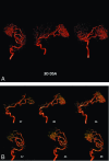
A, 3D-DSA of an AVM supplied by 2 branches of the right MCA. Despite the excellent image quality, the image is a composite of all of the 2D projections used in the reconstruction. The overlap of vascular components in the nidus precludes identification of intranidal aneurysms, the direct AVF, or venous outflow stenosis. B, Two views of early timeframes from a 4D-DSA reconstruction of the AVM shown in part A. The projections for this reconstruction are obtained at 30 frames per second. The number of each timeframe is shown beside each image. The angioarchitecture of the nidus can be clearly seen. The 4D-DSA images may be viewed from any desired angle at any time in the bolus passage.

References

    1. Davis B, Royalty K, Kowarschik M, et al. . 4D digital subtraction angiography: implementation and demonstration of feasibility. AJNR Am J Neuroradiol 2013;34:1914–21 10.3174/ajnr.A3529 - DOI - PMC - PubMed
    1. Sandoval-Garcia C, Royalty K, Yang P, et al. . 4D-DSA a new technique for arteriovenous malformation evaluation: a feasibility study. J Neurointerv Surg 2016;8:300–04 10.1136/neurintsurg-2014-011534 - DOI - PMC - PubMed
    1. Sandoval-Garcia C, Royalty K, Aagaard-Kienitz B, et al. . A comparison of 4D-DSA with 2D and 3D-DSA in the analysis of normal vascular structures in a canine model. AJNR Am J Neuroradiol 2015;36:1959–63 10.3174/ajnr.A4359 - DOI - PMC - PubMed
    1. Harris PA, Taylor R, Thielke R, et al. . Research electronic data capture (REDCap): a metadata-driven methodology and workflow process for providing translational research informatics support. J Biomed Inform 2009;42:377–81 10.1016/j.jbi.2008.08.010 - DOI - PMC - PubMed
    1. Fleiss JL. Measuring nominal scale agreement among many raters. Psychol Bull 1971;76:378–82 10.1037/h0031619 - DOI

Publication types

MeSH terms

LinkOut - more resources