. 2017 Jun;102(6):1054-1065.
doi: 10.3324/haematol.2016.159053. Epub 2017 Mar 9.

Targeted therapy for a subset of acute myeloid leukemias that lack expression of aldehyde dehydrogenase 1A1

Affiliations

Targeted therapy for a subset of acute myeloid leukemias that lack expression of aldehyde dehydrogenase 1A1

Maura Gasparetto et al. Haematologica. 2017 Jun.

Abstract

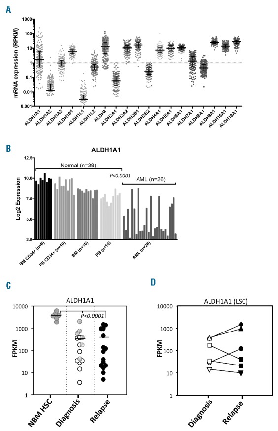
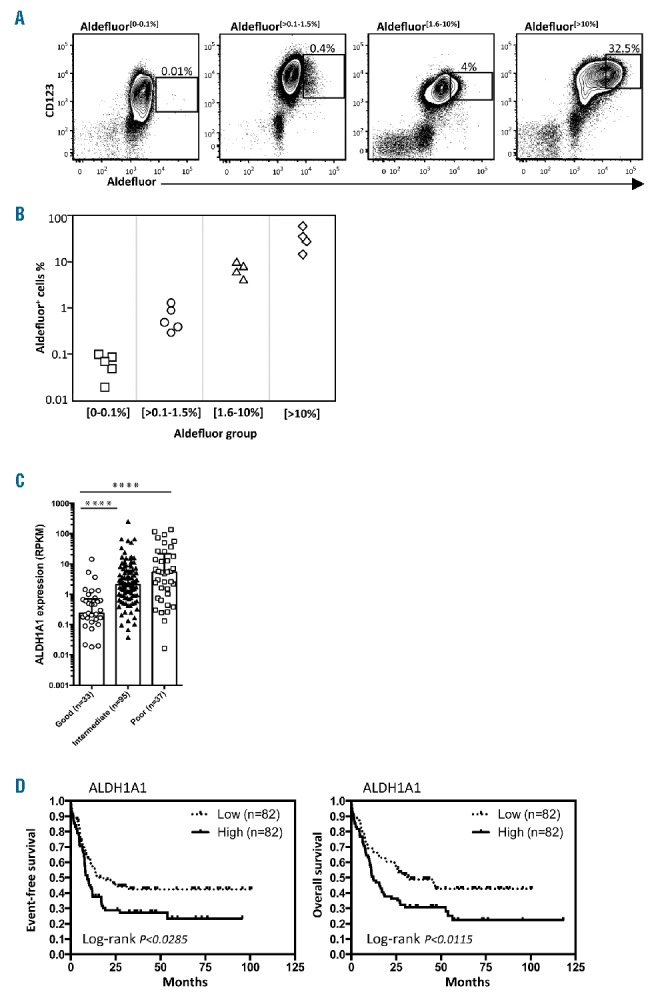
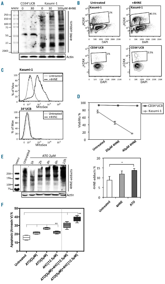
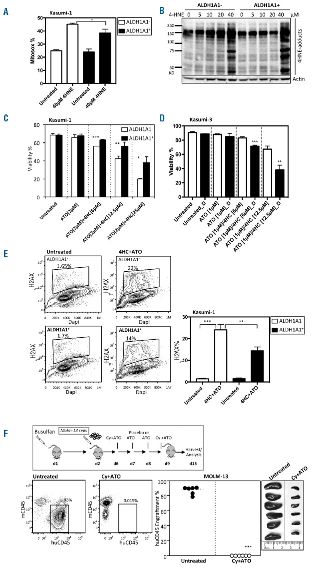
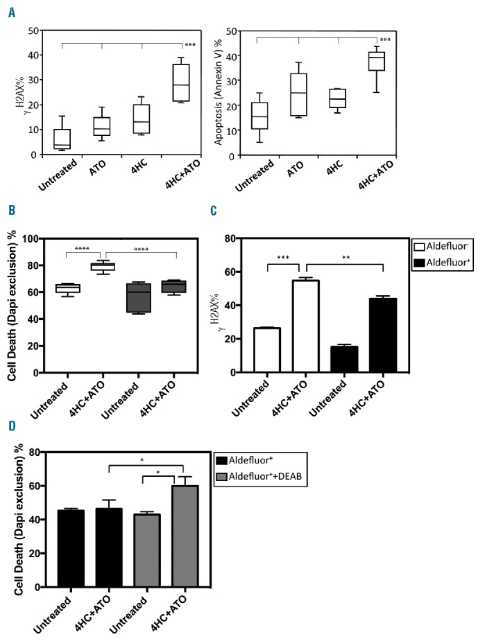
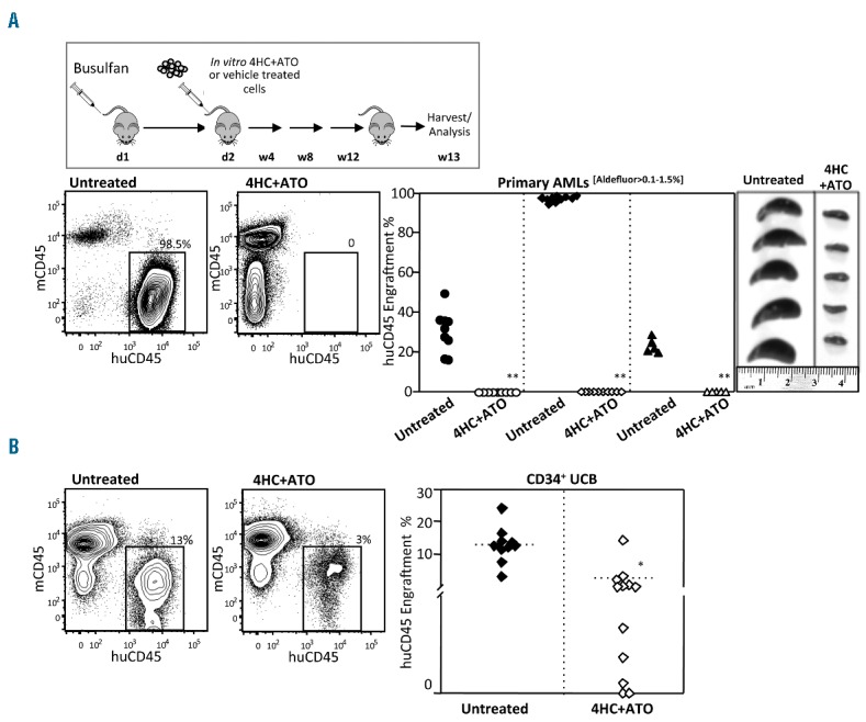
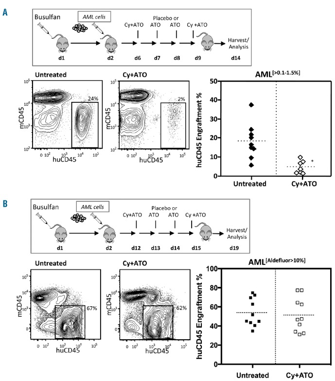
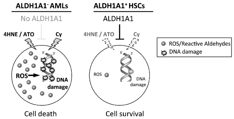
Aldehyde dehydrogenase 1A1 (ALDH1A1) activity is high in hematopoietic stem cells and functions in part to protect stem cells from reactive aldehydes and other toxic compounds. In contrast, we found that approximately 25% of all acute myeloid leukemias expressed low or undetectable levels of ALDH1A1 and that this ALDH1A1- subset of leukemias correlates with good prognosis cytogenetics. ALDH1A1- cell lines as well as primary leukemia cells were found to be sensitive to treatment with compounds that directly and indirectly generate toxic ALDH substrates including 4-hydroxynonenal and the clinically relevant compounds arsenic trioxide and 4-hydroperoxycyclophosphamide. In contrast, normal hematopoietic stem cells were relatively resistant to these compounds. Using a murine xenotransplant model to emulate a clinical treatment strategy, established ALDH1A1- leukemias were also sensitive to in vivo treatment with cyclophosphamide combined with arsenic trioxide. These results demonstrate that targeting ALDH1A1- leukemic cells with toxic ALDH1A1 substrates such as arsenic and cyclophosphamide may be a novel targeted therapeutic strategy for this subset of acute myeloid leukemias.


Figures

Figure 1.
Figure 1.
(A) Variable expression of ALDH1A1 and other aldehyde dehydrogenase (ALDH) isoforms in human acute myeloid leukemia (AML). The AML dataset from The Cancer Genome Atlas (TCGA) was analyzed for differential expression of ALDH isoforms in human AML blasts. (B) The GSE9476 dataset was analyzed for differential expression of ALDH1A1 between normal human hematopoietic cells (HSCs) and AML samples. (C) ALDH1A1 expression based on RNA-Seq analysis in LSC (white) and non-LSC (light gray) samples at diagnosis and LSC samples at relapse (black). The control is similarly analyzed normal bone marrow CD34+ cells (NBM HSC) (dark gray) (****P<0.0001). (D) ALDH1A1 expression using RNA-Seq analysis between paired diagnostic and relapsed LSC samples. (each symbol represents a different patient).
Figure 2.
Figure 2.
Levels of ALDH1A1 are variable between acute myeloid leukemias (AMLs) and have prognostic significance. (A) Aldefluor staining was performed to analyze single cell ALDH activity in representative samples of human primary AMLs that co-expressed CD123, a useful discriminator of AML and normal hematopoietic stem cells (HSCs). (B) Summary of Aldefluor staining in 18 different AML samples. (C) Association of ALDH1A1 expression level and poor, intermediate and good risk cytogenetic categories (TCGA dataset; ****P<0.0001). (D) Event-free survival (EFS) and overall survival (OS) Kaplan-Meier curve plots showing prognostic value of ALDH1A1 expression in all AML samples analyzed in the Cancer Genome Atlas dataset.
Figure 3.
Figure 3.
ALDH1A1 acute myeloid leukemia (AML) cell lines are sensitive to toxic substrates of aldehyde dehydrogenase (ALDH). (A) Western Blot measurement of 4HNE protein adducts in Kasumi-1 and normal CD34+ umbilical cord blood (UCB) cells treated with 30 μM 4HNE or vehicle. Kasumi-1 cells treated with 100 μM 4HNE were included as a positive control. (B) Flow cytometric assessment of DNA damage (γH2AX) in Kasumi-1 with and without 4HNE treatment compared to normal CD34+ UCB cells. (C) Flow cytometric assessment of ROS in Kasumi-1 with and without 30–40 μM 4HNE treatment compared to normal CD34+ UCB cells. (D) 4HNE dose response curves in Kasumi-1 and normal CD34+ cells treated with 20 and 40 μM 4HNE [10 μM 4HNE was added to cell media every hour, data is mean and standard deviation (SD) of triplicate samples]. (E) Western blot analysis of 4HNE protein adducts generated by treating Kasumi-1 cells with 2 μM ATO over 12 hours and flow cytometric analysis of 4HNE adducts in Kasumi-1 induced by 2.5 μM 4HNE and 2 μM ATO treatment at 12 hours (n=3; *P<0.05). (F) Apoptosis of Kasumi-1 cells following overnight treatment with ATO (2 μM or 5 μM) or 4HC (12.5 μM) alone and in combination (n=6 replicates; **P<0.0001). (D–F) Bars represent mean±SD. (D and E) Results of technical triplicates are shown.
Figure 4.
Figure 4.
Overexpression of ALDH1A1+ partially rescues ALDH1A1 acute myeloid leukemia (AML) cell sensitivity to toxic substrates of ALDH. (A) ROS (Mitosox) detection in ALDH1A1+ and ALDH1A1- Kasumi-1 cells after six hours of treatment with 40 μM 4-HNE (n=3;*P<0.05). (B) Western blot analysis of 4-HNE protein adducts generated by treating ALDH1A1+ and ALDH1A1 Kasumi-1 cells with different concentrations of 4-HNE for 1 hour. (C) Viability of Kasumi-1 cells engineered to express ALDH1A1 through lentiviral gene transfer and treated with various combinations and doses of 4HC and ATO (n=3 replicates; ***P<0.0005, **P<0.005, *P<0.05). (D) Viability of Kasumi-3 cells treated with various combinations and doses of 4HC and ATO with (black) or without (white) the ALDH inhibitor DEAB (D) (n=3; **P<0.005, ***P<0.0005). (E) Flow cytometric assessment and summary graph of DNA damage (γH2AX) in ALDH1A1+ and ALDH1A1 Kasumi-1 cells treated with 4HC+ATO (n=3; **P<0.005, ***P<0.0005). (A–C) Bars represent mean±SD. Technical triplicates were performed. (F) In vivo treatment with Cy and ATO of NSGS mice with established ALDH1A1 MOLM-13 leukemia (n=6; ***P<0.0005).
Figure 5.
Figure 5.
Primary human ALDH1A1 acute myeloid leukemias (AMLs) are sensitive to 4HC+ATO while ALDH1A1+ AMLs are relatively resistant. (A) In vitro sensitivity of primary human AMLs to 4HC and ATO. Primary human AMLs were treated overnight with 4HC+ATO (12.5 μM+2 μM) and DNA damage and cell viability were measured (4 AMLs in triplicate; ***P<0.0005). (B) Sensitivity of Aldefluor+ and Aldefluor fractions from 2 representative AMLs purified by FACS and then treated overnight with 4HC+ATO (12.5 μM+2 μM) (n=6; **P<0.005, ***P<0.0005). (C) DNA damage (γH2AX) of Aldefluor+ and Aldefluor fractions from an ALDH1A1+ AML following overnight treatment with 4HC+ATO (n=3; **P<0.005, ***P<0.0005). (D) Relative sensitivity of Aldefluor+ fraction from an ALDH1A1+ AML to 4HC+ATO with and without 5 μM DEAB at 18 hours (n=3; *P<0.05). (A–D) Bars represent mean±SD. (C–D) Results of technical triplicates are shown.
Figure 6.
Figure 6.
ALDH1A1 acute myeloid leukemias (AMLs) are sensitive to 4HC+ATO while normal HSCs are relatively resistant. (A) 4HC+ATO in vitro treatment eliminated engraftment of primary AMLs in NSGS mice. Representative flow cytometric analysis of harvested bone marrow of Aldefluor[0–0.1%] AMLs that underwent overnight ex vivo treatment with 4HC+ATO (30 μM+ 5 μM) or vehicle (left panels) and summary of xenotransplant data from the 3 AMLs treated in vitro and transplanted into NSGS mice (right panels). The data are a summary of 25 of 30 control and 25 of 30 treated mice analyzed after 12 weeks (**P<0.0005). (B) In contrast, 4HC+ATO in vitro treatment did not completely eliminate engraftment of normal CD34+UCB cells in NSGS mice. NSGS mice transplanted with 2 pooled UCBs following 4HC+ATO or vehicle treatment as above (treated: n=12; untreated: n=10). While level of engraftment was reduced, in contrast to the AML experiments above, 7 of 12 mice treated with normal CD34+UCB cells displayed more than 1% engraftment (*P<0.005). Analysis of engraftment in bone marrow was performed at 13 weeks using flow cytometry.
Figure 7.
Figure 7.
ALDH1A1 acute myeloid leukemias (AMLs) are sensitive to Cy+ATO while ALDH1A1+ AMLs are relatively resistant. (A) In vivo Cy+ATO treatment reduced leukemic burden in NSGS mice engrafted with primary ALDH1A1 AML cells (treated: n=7; untreated: n=9; *P<0.005). (B) In vivo Cy+ATO treatment shows no differences in leukemic burden in NSGS mice engrafted with primary ALDH1A1+ AML cells (n=10; not significant).
Figure 8.
Figure 8.
Model for selectively targeting ALDH1A1 acute myeloid leukemias (AMLs). In this treatment strategy, AMLs will be profiled for aldehyde dehydrogenase (ALDH) isoform expression and those lacking ALDH1A1 expression will be treated with agents that directly and indirectly generate lethal levels of ALDH substrates including ROS, reactive aldehydes and others. In contrast, normal hematopoietic stem cells express high levels of ALDH1A1 that could metabolize these compounds resulting in relative sparing.

Similar articles

Cited by

References

    1. Vasiliou V, Thompson DC, Smith C, Fujita M, Chen Y. Aldehyde dehydrogenases: from eye crystallins to metabolic disease and cancer stem cells. Chem Biol Interact. 2013;202(1–3):2–10. - PMC - PubMed
    1. Singh S, Brocker C, Koppaka V, et al. Aldehyde dehydrogenases in cellular responses to oxidative/electrophilic stress. Free Radic Biol Med. 2013;56:89–101. - PMC - PubMed
    1. Storms RW, Trujillo AP, Springer JB, et al. Isolation of primitive human hematopoietic progenitors on the basis of aldehyde dehydrogenase activity. Proc Natl Acad Sci USA. 1999;96(16):9118–9123. - PMC - PubMed
    1. Gasparetto M, Sekulovic S, Brocker C, et al. Aldehyde dehydrogenases are regulators of hematopoietic stem cell numbers and B-cell development. Exp Hematol. 2012; 40(4):318–329.e312. - PubMed
    1. Levi BP, Yilmaz OH, Duester G, Morrison SJ. Aldehyde dehydrogenase 1a1 is dispensable for stem cell function in the mouse hematopoietic and nervous systems. Blood. 2009;113(8):1670–1680. - PMC - PubMed