

Dynamics of middle cerebral artery blood flow velocity during moderate-intensity exercise
- PMID: 28280106
- PMCID: PMC5451537
- DOI: 10.1152/japplphysiol.00995.2016
Dynamics of middle cerebral artery blood flow velocity during moderate-intensity exercise
Abstract
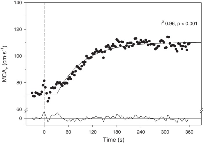
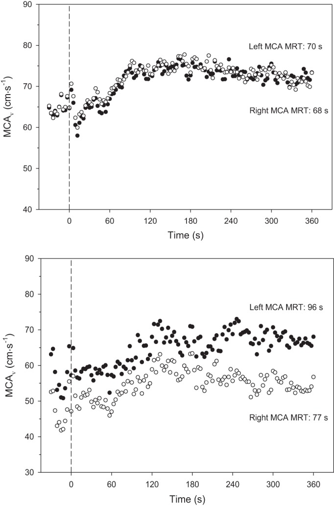
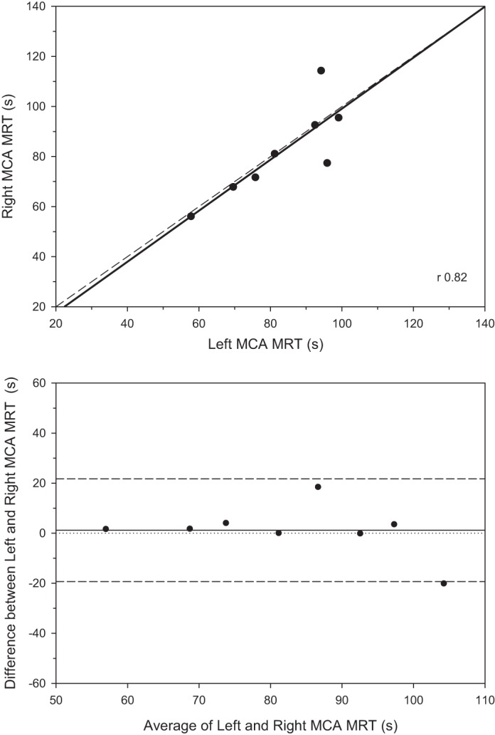
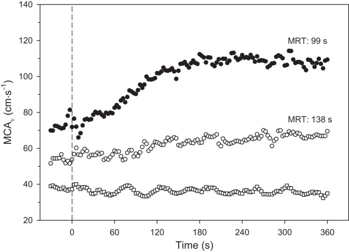
The dynamic response to a stimulus such as exercise can reveal valuable insights into systems control in health and disease that are not evident from the steady-state perturbation. However, the dynamic response profile and kinetics of cerebrovascular function have not been determined to date. We tested the hypotheses that bilateral middle cerebral artery blood flow mean velocity (MCAV) increases exponentially following the onset of moderate-intensity exercise in 10 healthy young subjects. The MCAV response profiles were well fit to a delay (TD) + exponential (time constant, τ) model with substantial agreement for baseline [left (L): 69, right (R): 64 cm/s, coefficient of variation (CV) 11%], response amplitude (L: 16, R: 13 cm/s, CV 23%), TD (L: 54, R: 52 s, CV 9%), τ (L: 30, R: 30 s, CV 22%), and mean response time (MRT) (L: 83, R: 82 s, CV 8%) between left and right MCAV as supported by the high correlations (e.g., MRT r = 0.82, P < 0.05) and low CVs. Test-retest reliability was high with CVs for the baseline, amplitude, and MRT of 3, 14, and 12%, respectively. These responses contrasted markedly with those of three healthy older subjects in whom the MCAV baseline and exercise response amplitude were far lower and the kinetics slowed. A single older stroke patient showed baseline ipsilateral MCAV that was lower still and devoid of any exercise response whatsoever. We conclude that kinetics analysis of MCAV during exercise has significant potential to unveil novel aspects of cerebrovascular function in health and disease.NEW & NOTEWORTHY Resolution of the dynamic stimulus-response profile provides a greater understanding of the underlying the physiological control processes than steady-state measurements alone. We report a novel method of measuring cerebrovascular blood velocity (MCAv) kinetics under ecologically valid conditions from rest to moderate-intensity exercise. This technique reveals that brain blood flow increases exponentially following the onset of exercise with 1) a strong bilateral coherence in young healthy individuals, and 2) a potential for unique age- and disease-specific profiles.
Keywords: aging; blood flow velocity; brain blood flow; middle cerebral artery; stroke.
Copyright © 2017 the American Physiological Society.
Figures
References
-
- Alexandrov AV, Sloan MA, Wong LK, Douville C, Razumovsky AY, Koroshetz WJ, Kaps M, Tegeler CH; American Society of Neuroimaging Practice Guidelines Committee . Practice standards for transcranial Doppler ultrasound: part I–test performance. J Neuroimaging 17: 11–18, 2007. doi: 10.1111/j.1552-6569.2006.00088.x. - DOI - PubMed
MeSH terms
Grants and funding
LinkOut - more resources
Full Text Sources
Other Literature Sources
Medical