

Oxidative stress elicited by modifying the ceramide acyl chain length reduces the rate of clathrin-mediated endocytosis
- PMID: 28280117
- PMCID: PMC5399788
- DOI: 10.1242/jcs.199968
Oxidative stress elicited by modifying the ceramide acyl chain length reduces the rate of clathrin-mediated endocytosis
Abstract
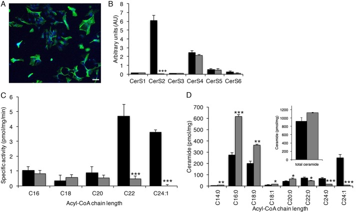
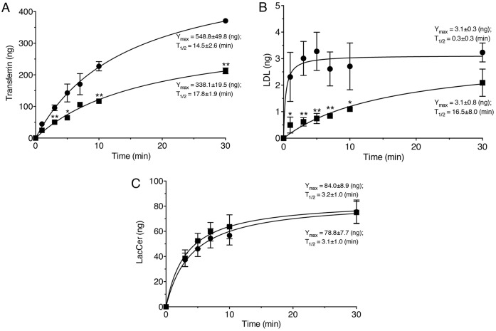
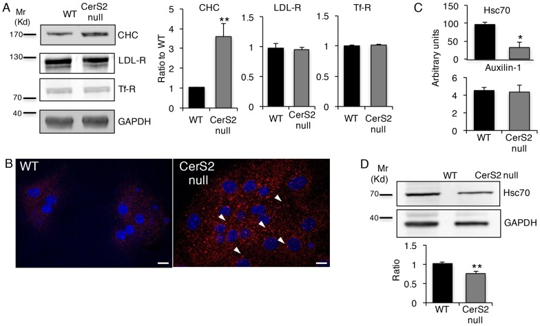
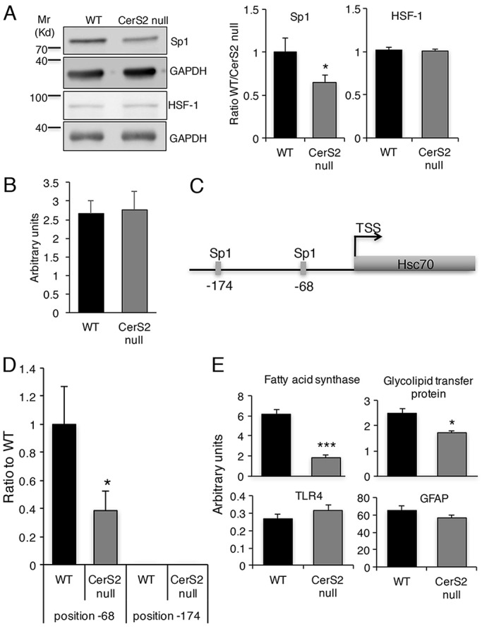
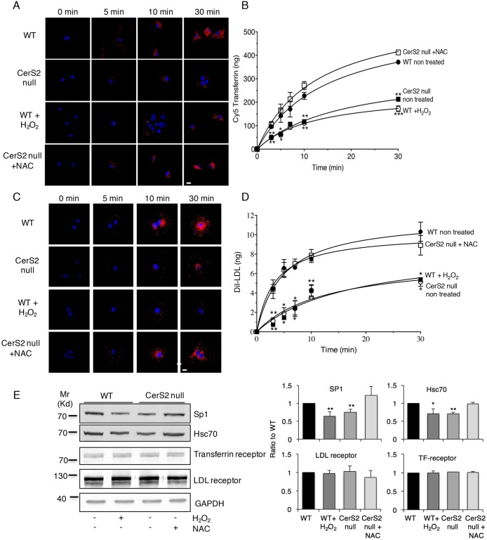
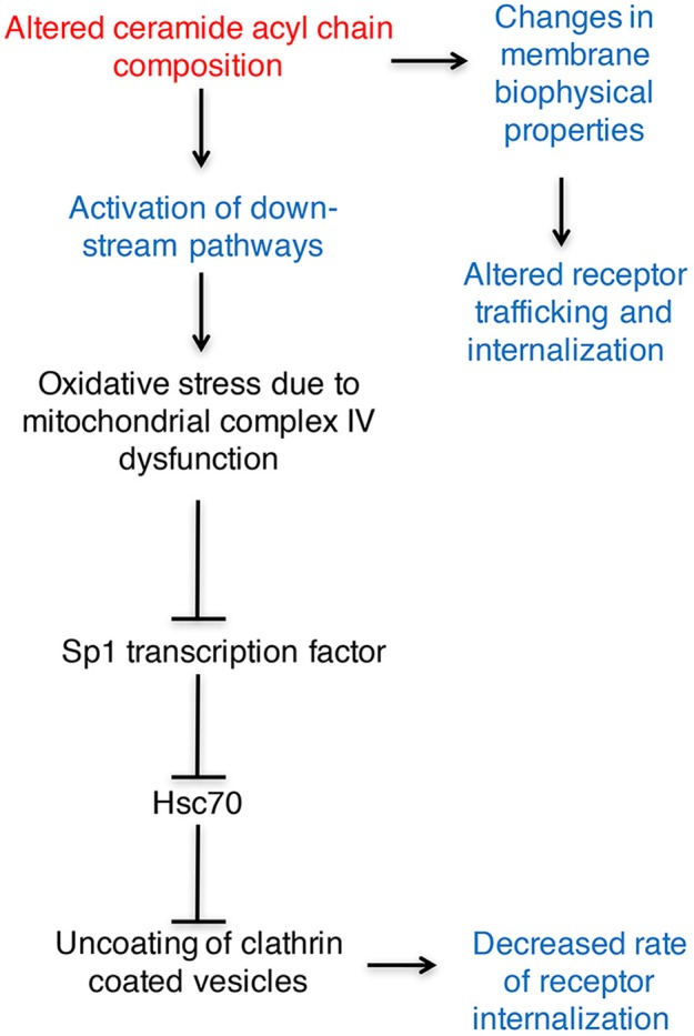
Sphingolipids modulate clathrin-mediated endocytosis (CME) by altering the biophysical properties of membranes. We now examine CME in astrocytes cultured from ceramide synthase 2 (CerS2) null mice, which have an altered sphingolipid acyl chain composition. The rate of endocytosis of low-density lipoprotein and transferrin, which are internalized via CME, was reduced in CerS2 null astrocytes, although the rate of caveolin-mediated endocytosis was unaltered. Levels of clathrin heavy chain were increased, which was due to decreased levels of Hsc70 (also known as HSPA8), a protein involved in clathrin uncoating. Hsc70 levels were decreased because of lower levels of binding of Sp1 to position -68 in the Hsc70 promoter. Levels of Sp1 were downregulated due to oxidative stress, which was elevated fourfold in CerS2 null astrocytes. Furthermore, induction of oxidative stress in wild-type astrocytes decreased the rate of CME, whereas amelioration of oxidative stress in CerS2 null astrocytes reversed the decrease. Our data are consistent with the notion that sphingolipids not only change membrane biophysical properties but also that changes in their composition can result in downstream effects that indirectly impinge upon a number of cellular pathways, such as CME.
Keywords: Clathrin-mediated endocytosis; Hsc70; Reactive oxygen species; Sp1; Very long-chain ceramides.
© 2017. Published by The Company of Biologists Ltd.
Conflict of interest statement
The authors declare no competing or financial interests.
Figures

Similar articles
-
Altering sphingolipid composition with aging induces contractile dysfunction of gastric smooth muscle via K(Ca) 1.1 upregulation.Aging Cell. 2015 Dec;14(6):982-94. doi: 10.1111/acel.12388. Epub 2015 Aug 20. Aging Cell. 2015. PMID: 26288989 Free PMC article.
-
Ablation of ceramide synthase 2 causes chronic oxidative stress due to disruption of the mitochondrial respiratory chain.J Biol Chem. 2013 Feb 15;288(7):4947-56. doi: 10.1074/jbc.M112.402719. Epub 2013 Jan 2. J Biol Chem. 2013. PMID: 23283968 Free PMC article.
-
Hepatic fatty acid uptake is regulated by the sphingolipid acyl chain length.Biochim Biophys Acta. 2014 Dec;1841(12):1754-66. doi: 10.1016/j.bbalip.2014.09.009. Biochim Biophys Acta. 2014. PMID: 25241943 Free PMC article.
-
The role of ceramide in regulating endoplasmic reticulum function.Biochim Biophys Acta Mol Cell Biol Lipids. 2020 Jan;1865(1):158489. doi: 10.1016/j.bbalip.2019.06.015. Epub 2019 Jun 21. Biochim Biophys Acta Mol Cell Biol Lipids. 2020. PMID: 31233888 Review.
-
The effect of altered sphingolipid acyl chain length on various disease models.Biol Chem. 2015 Jun;396(6-7):693-705. doi: 10.1515/hsz-2014-0310. Biol Chem. 2015. PMID: 25720066 Review.
Cited by
-
Ceramide and Related Molecules in Viral Infections.Int J Mol Sci. 2021 May 26;22(11):5676. doi: 10.3390/ijms22115676. Int J Mol Sci. 2021. PMID: 34073578 Free PMC article. Review.
-
Endocytosis in the adaptation to cellular stress.Cell Stress. 2020 Aug 18;4(10):230-247. doi: 10.15698/cst2020.10.232. Cell Stress. 2020. PMID: 33024932 Free PMC article. Review.
-
Neutral Ceramidase Is Required for the Reproduction of Brown Planthopper, Nilaparvata lugens (Stål).Front Physiol. 2021 Feb 24;12:629532. doi: 10.3389/fphys.2021.629532. eCollection 2021. Front Physiol. 2021. PMID: 33716775 Free PMC article.
-
Eleven residues determine the acyl chain specificity of ceramide synthases.J Biol Chem. 2018 Jun 22;293(25):9912-9921. doi: 10.1074/jbc.RA118.001936. Epub 2018 Apr 9. J Biol Chem. 2018. PMID: 29632068 Free PMC article.
-
Agonists that stimulate secretion promote the recruitment of CFTR into membrane lipid microdomains.J Gen Physiol. 2019 Jun 3;151(6):834-849. doi: 10.1085/jgp.201812143. Epub 2019 May 2. J Gen Physiol. 2019. PMID: 31048413 Free PMC article.
References
Publication types
MeSH terms
Substances
Grants and funding
LinkOut - more resources
Full Text Sources
Other Literature Sources
Molecular Biology Databases
Miscellaneous
