. 2017 Apr 3;216(4):1035-1049.
doi: 10.1083/jcb.201611088. Epub 2017 Mar 9.

Tango1 spatially organizes ER exit sites to control ER export

Affiliations

Tango1 spatially organizes ER exit sites to control ER export

Min Liu et al. J Cell Biol. .

Abstract

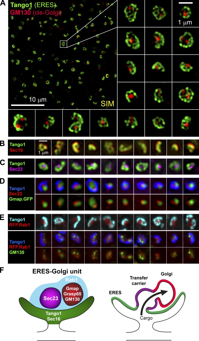
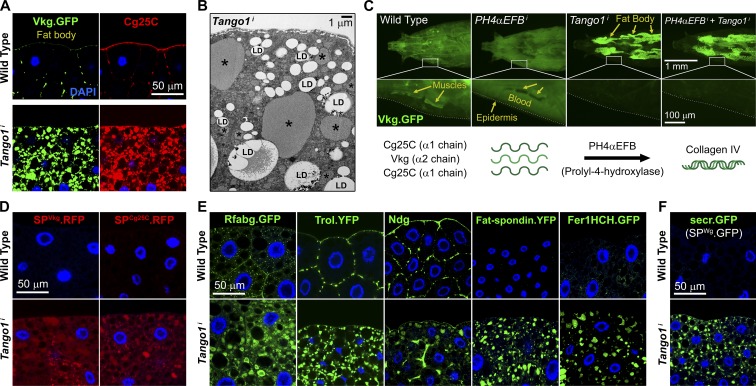
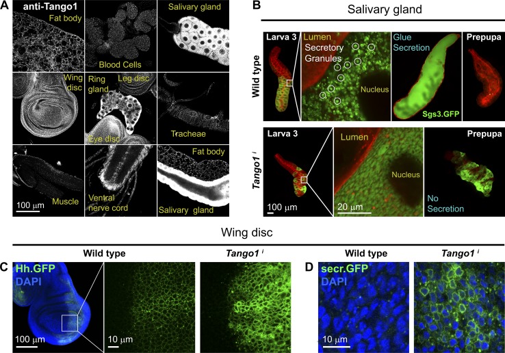
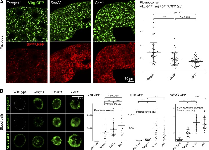
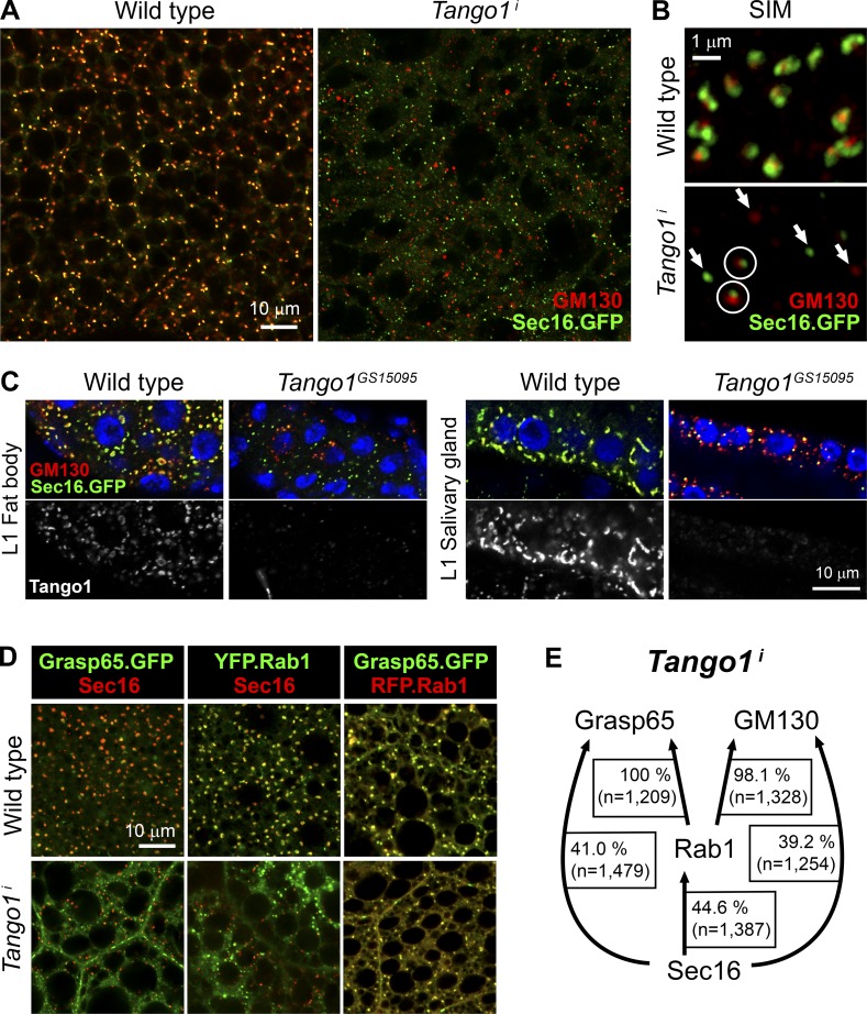
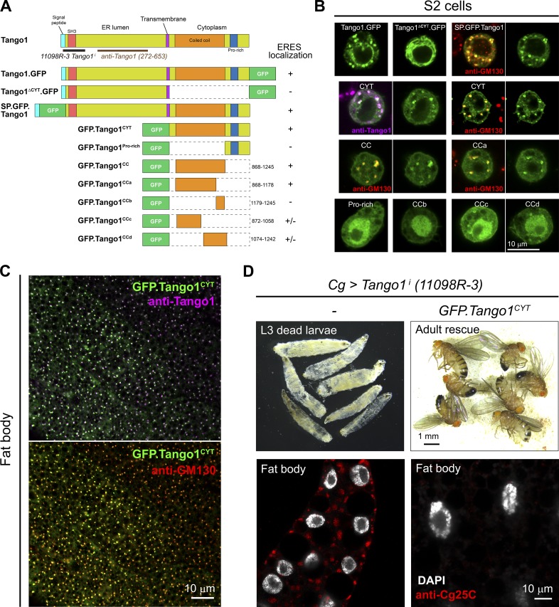
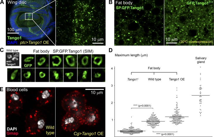
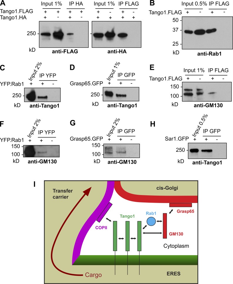
Exit of secretory cargo from the endoplasmic reticulum (ER) takes place at specialized domains called ER exit sites (ERESs). In mammals, loss of TANGO1 and other MIA/cTAGE (melanoma inhibitory activity/cutaneous T cell lymphoma-associated antigen) family proteins prevents ER exit of large cargoes such as collagen. Here, we show that Drosophila melanogaster Tango1, the only MIA/cTAGE family member in fruit flies, is a critical organizer of the ERES-Golgi interface. Tango1 rings hold COPII (coat protein II) carriers and Golgi in close proximity at their center. Loss of Tango1, present at ERESs in all tissues, reduces ERES size and causes ERES-Golgi uncoupling, which impairs secretion of not only collagen, but also all other cargoes we examined. Further supporting an organizing role of Tango1, its overexpression creates more and larger ERESs. Our results suggest that spatial coordination of ERES, carrier, and Golgi elements through Tango1's multiple interactions increases secretory capacity in Drosophila and allows secretion of large cargo.


Figures

Figure 1.
Figure 1.
Imaging of ERES–Golgi units through SIM microscopy. (A) Image of L3 fat body stained with anti-Tango1 and anti-GM130 antibodies obtained through super-resolution SIM imaging. Examples of individual ERES–Golgi units are shown at higher magnification. Images are maximum intensity projections of two to five confocal sections. (B) ERESs visualized through staining with anti-Sec16 (ERES marker) and anti-Tango1. (C) ERES visualized through staining with anti-Sec23 (COPII coat) and anti-Tango1. (D) ERES–Golgi units visualized with anti-Tango1, anti-Sec23, and Gmap.GFP (GFP-trap insertion into Gmap gene). (E) Localization of Rab1 (Cg>RFP.Rab1) in relation to ERESs (anti-Tango1) and cis-Golgi (anti-GM130). (F) Organization of ERES–Golgi units as deduced from SIM images above (A–E).
Figure 2.
Figure 2.
Tango1 knockdown impairs general secretion in fat body cells. (A) Confocal images of fat body cells from wild-type (top) and Tango1 knockdown (BM-40-SPARC-GAL4 > UAS-Tango1i, bottom) third-instar larvae (L3 stage) showing distribution of Collagen IV chains α2 (Vkg.GFP, left) and α1 (anti-Cg25C, right). Nuclei were stained with DAPI. (B) Electron micrograph of Cg>Tango1i fat body. Asterisks mark cargo-filled dilated ER. LD, lipid droplets. (C) Distribution of Collagen IV (Vkg.GFP) in wild-type, BM-40-SPARC>PH4αEFBi, >Tango1i, and double >PH4αEFBi+Tango1i larvae. Insets magnified in the bottom panels show Vkg.GFP localization to muscle basement membranes (wild type) or diffuse blood signal (PH4αEFBi). (D) Wild-type (top) and Tango1i (bottom) fat body cells expressing RFP coupled to signal peptides of Vkg (Cg>SPVkg.RFP, left) and Cg25C (Cg>SPCg25C.RFP, right). (E) Wild-type and Tango1i fat body cells showing distribution of Rfabg (Apo-B, Rfabg.sGFPfTRG.900), YFP-tagged Trol (Perlecan, trolCPTI-002049.YFP), Nidogen (anti-Ndg staining), Fat-spondin (fat-spondinCPTI001685.YFP), and Ferritin (Fer1HCHG188.GFP). (F) Wild-type and Tango1i fat body cells expressing secr.GFP (BM-40-SPARC>secr.GFP, signal peptide of Wingless coupled to GFP).
Figure 3.
Figure 3.
Tango1 is widely expressed and required for secretion in salivary glands and disc cells. (A) Expression of Tango1 in different tissues of the L3 larva (anti-Tango1 staining). (B) Localization of glue protein Sgs3 (Sgs3.GFP) in late L3 and pre-pupal salivary glands of wild-type and He>Tango1i animals. No individualized secretory granules are seen in Tango1i glands, and Sgs3.GFP remains inside cells after the onset of metamorphosis. The membrane marker myr.RFP is shown in red. (C) Confocal images of control and Tango1i wing discs expressing Hedgehog.GFP in their posterior compartment (hh>hh.GFP). (D) Confocal images of wild-type and Tango1i wing disc cells expressing secr.GFP (rn>secr.GFP).
Figure 4.
Figure 4.
Tango1 differentially affects the secretion of Collagen IV. (A) Confocal images of fat body cells from BM-40-SPARC>Tango1i, >Sec23i, and >Sar1i larvae showing distribution of Collagen IV.GFP (Vkg.GFP) and RFP (signal peptide of Vkg coupled to RFP). Graphs show quantification of fluorescent signal ratio, each dot representing an individual cell (n > 50). Horizontal lines and error bars represent mean and standard deviation, respectively. P-values correspond to two-tailed t tests. au, arbitrary units. (B) Confocal images of hemocytes (blood cells) from wild-type, BM-40-SPARC>Tango1i, >Sec23i, and >Sar1i larvae showing distribution of Vkg.GFP (top), secr.GFP (middle), and VSVG.GFP (bottom). Graphs show quantification of fluorescent signal as above (A; n ≥ 22). au, arbitrary units.
Figure 5.
Figure 5.
Loss of Tango1 produces smaller ERESs uncoupled from Golgi. (A) Confocal images of wild-type and Cg>Tango1i fat body cells stained with anti-GM130 and expressing Sec16.GFP (transgenic insertion of BAC construct containing Sec16 locus modified by the addition of C-terminal GFP). (B) SIM images of wild-type and Cg>Tango1i fat body showing localization of Sec16.GFP and GM130. Arrows mark separate Sec16- and GM130-positive structures. The two structures where Sec16- and GM130-positive structures maintain proximity are marked by circles. (C) Confocal images of fat body (left) and salivary gland (right) cells from wild-type and mutant Tango1GS15095 first instar (L1) larvae showing localization of Sec16.GFP and GM130. Tissues were stained with anti-Tango1 antibody. Nuclei are in blue (DAPI). (D) Confocal images of fat body from wild-type (top) and Cg>Tango1i (bottom) larvae showing localization of the cis-Golgi marker Grasp65 (Cg>Grasp65.GFP), the ERES marker Sec16, and the GTPase Rab1 (Cg>YFP.Rab1 and Cg>RFP.Rab1). (E) Quantification of defects in ERES–Golgi unit organization in Cg>Tango1i fat body cells. The percentage of Sec16 puncta that are found next to Rab1, Grasp65, and GM130 puncta is indicated. Idem for Rab1-positive puncta next to Grasp65 and GM130 puncta.
Figure 6.
Figure 6.
The cytoplasmic part of Tango1 directs ERES localization and can rescue Tango1 loss. (A) Graphic representation of GFP-tagged Tango1 constructs generated in this study. The ability to localize to ERESs is indicated on the right. (B) Confocal images of Drosophila S2 cells transfected with the indicated constructs. GM130 and Tango1 antibody staining used to confirm ERES–Golgi localization. (C) Localization of Tango1CYT (Cg>GFP.Tango1CYT) in fat body cells stained with anti-Tango1 and anti-GM130. Note that Tango1 antibody was raised against a part of Tango1 not present in Tango1CYT. (D) Rescue of Tango1i larval lethality and Collagen IV retention by GFP.Tango1CYT. See also Table S2. Note that the double-stranded RNA used here targets a part of the Tango1 sequence not present in Tango1CYT.
Figure 7.
Figure 7.
Tango1 overexpression increases ERES size and number. (A) Confocal images of a wing disc overexpressing (OE) Tango1 in a medial stripe of cells (ptc>Tango1) and stained with anti-Tango1. Area inside the square is shown at higher magnification on the right. (B) Confocal images of fat body expressing Tango1 GFP-tagged at the N terminus after its signal peptide (Cg>SP.GFP.Tango1, left) and the cytoplasmic portion of Tango1 GFP-tagged at the N terminus (GFP.Tango1CYT, right). (C) Examples of enlarged ERESs caused by expression of SP.GFP.Tango1 imaged through SIM. Two normal-sized wild-type ERESs are shown for comparison. (D) Quantification of individual ERES size in Cg>Tango1i fat body (n = 202), wild-type fat body (n = 163), Tango1-overexpressing fat body (Cg>SP.GFP.Tango1; n = 174), and wild-type salivary glands (n = 43). Horizontal lines indicate mean size. Images were analyzed with ImageJ, and apparent maximum lengths of the structures were manually measured with the line tool. P-values from two-tailed t tests are shown. (E) Confocal images of blood cells from wild-type (left) and Cg>SP.GFP.Tango1 larvae (right) stained with anti-Gmap (cis-Golgi marker).
Figure 8.
Figure 8.
Multiple interactions of Tango1 at the ERES–Golgi interface. (A) Tango1.HA (left) and Tango1.FLAG (right) were immunoprecipitated from L3 fat body lysates. Immunoprecipitates (IP) were Western blotted with anti-FLAG (left) and anti-HA (right) antibodies. (B) Tango1.FLAG fat body immunoprecipitates, Western blotted with anti-Rab1 (see Fig. S4 for antibody validation). (C) YFP.Rab1 fat body immunoprecipitates, Western blotted with anti-Tango1. (D) Grasp65.GFP fat body immunoprecipitates, Western blotted with anti-Tango1. (E) Tango1.FLAG fat body immunoprecipitates, Western blotted with anti-GM130. (F) YFP.Rab1 fat body immunoprecipitates, Western blotted with anti-GM130. (G) Grasp65.GFP fat body immunoprecipitates, Western blotted with anti-GM130. (H) Sar1.GFP fat body immunoprecipitates, Western blotted with anti-Tango1. (I) Static model depicting Tango1 interactions (with itself, COPII, Rab1, and cis-Golgi peripheral proteins GM130-Grasp65) and possible role of Tango1 in maintaining Golgi proximity and ERES–Golgi organization for increased secretory capacity.

Similar articles

Cited by

References

    1. Abrams E.W., and Andrew D.J.. 2005. CrebA regulates secretory activity in the Drosophila salivary gland and epidermis. Development. 132:2743–2758. 10.1242/dev.01863 - DOI - PubMed
    1. Bannykh S.I., Rowe T., and Balch W.E.. 1996. The organization of endoplasmic reticulum export complexes. J. Cell Biol. 135:19–35. 10.1083/jcb.135.1.19 - DOI - PMC - PubMed
    1. Bard F., Casano L., Mallabiabarrena A., Wallace E., Saito K., Kitayama H., Guizzunti G., Hu Y., Wendler F., Dasgupta R., et al. . 2006. Functional genomics reveals genes involved in protein secretion and Golgi organization. Nature. 439:604–607. 10.1038/nature04377 - DOI - PubMed
    1. Bharucha N., Liu Y., Papanikou E., McMahon C., Esaki M., Jeffrey P.D., Hughson F.M., and Glick B.S.. 2013. Sec16 influences transitional ER sites by regulating rather than organizing COPII. Mol. Biol. Cell. 24:3406–3419. 10.1091/mbc.E13-04-0185 - DOI - PMC - PubMed
    1. Bonifacino J.S., and Glick B.S.. 2004. The mechanisms of vesicle budding and fusion. Cell. 116:153–166. 10.1016/S0092-8674(03)01079-1 - DOI - PubMed

Publication types

MeSH terms

Substances

LinkOut - more resources