. 2017 Sep;58(9):1386-1394.
doi: 10.2967/jnumed.116.187310. Epub 2017 Mar 9.

89Zr-DFO-AMG102 Immuno-PET to Determine Local Hepatocyte Growth Factor Protein Levels in Tumors for Enhanced Patient Selection

Affiliations

89Zr-DFO-AMG102 Immuno-PET to Determine Local Hepatocyte Growth Factor Protein Levels in Tumors for Enhanced Patient Selection

Eric W Price et al. J Nucl Med. 2017 Sep.

Abstract

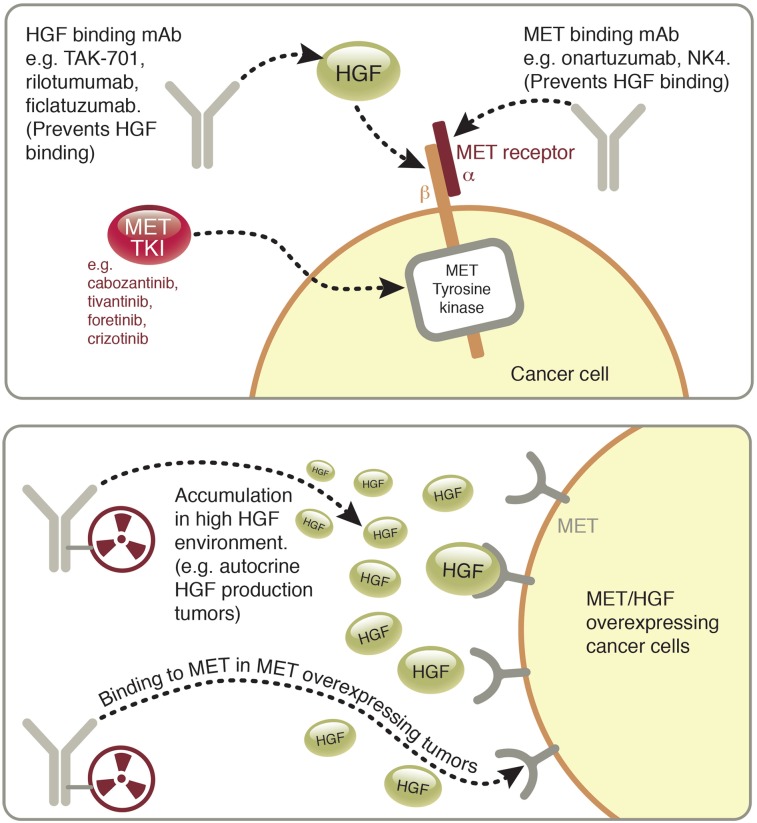
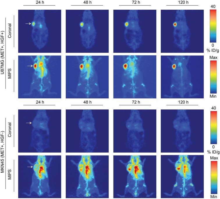
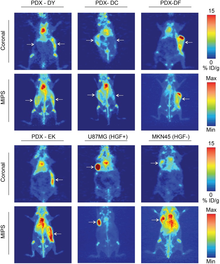
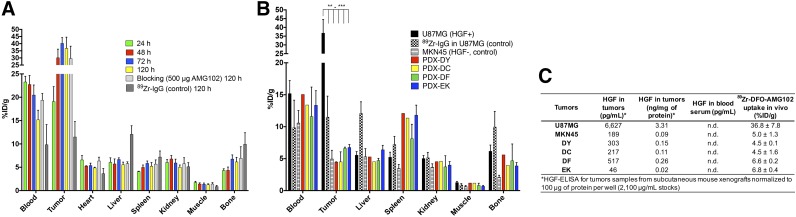
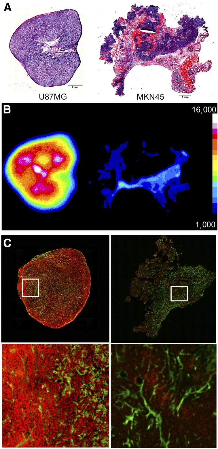
The hepatocyte growth factor (HGF) binding antibody rilotumumab (AMG102) was modified for use as a 89Zr-based immuno-PET imaging agent to noninvasively determine the local levels of HGF protein in tumors. Because recent clinical trials of HGF-targeting therapies have been largely unsuccessful in several different cancers (e.g., gastric, brain, lung), we have synthesized and validated 89Zr-DFO-AMG102 as a companion diagnostic for improved identification and selection of patients having high local levels of HGF in tumors. To date, patient selection has not been performed using the local levels of HGF protein in tumors. Methods: The chelator p-SCN-Bn-DFO was conjugated to AMG102, radiolabeling with 89Zr was performed in high radiochemical yields and purity (>99%), and binding affinity of the modified antibody was confirmed using an enzyme-linked immunosorbent assay (ELISA)-type binding assay. PET imaging, biodistribution, autoradiography and immunohistochemistry, and ex vivo HGF ELISA experiments were performed on murine xenografts of U87MG (HGF-positive, MET-positive) and MKN45 (HGF-negative, MET-positive) and 4 patient-derived xenografts (MET-positive, HGF unknown). Results: Tumor uptake of 89Zr-DFO-AMG102 at 120 h after injection in U87MG xenografts (HGF-positive) was high (36.8 ± 7.8 percentage injected dose per gram [%ID/g]), whereas uptake in MKN45 xenografts (HGF-negative) was 5.0 ± 1.3 %ID/g and a control of nonspecific human IgG 89Zr-DFO-IgG in U87MG tumors was 11.5 ± 3.3 %ID/g, demonstrating selective uptake in HGF-positive tumors. Similar experiments performed in 4 different gastric cancer patient-derived xenograft models showed low uptake of 89Zr-DFO-AMG102 (∼4-7 %ID/g), which corresponded with low HGF levels in these tumors (ex vivo ELISA). Autoradiography, immunohistochemical staining, and HGF ELISA assays confirmed that elevated levels of HGF protein were present only in U87MG tumors and that 89Zr-DFO-AMG102 uptake was closely correlated with HGF protein levels in tumors. Conclusion: The new immuno-PET imaging agent 89Zr-DFO-AMG102 was successfully synthesized, radiolabeled, and validated in vitro and in vivo to selectively accumulate in tumors with high local levels of HGF protein. These results suggest that 89Zr-DFO-AMG102 would be a valuable companion diagnostic tool for the noninvasive selection of patients with elevated local concentrations of HGF in tumors for planning any HGF-targeted therapy, with the potential to improve clinical outcomes.

Keywords: AMG102; HGF; MET; PET; hepatocyte growth factor; patient-derived xenograft; rilotumumab; scatter factor.


Figures

FIGURE 1.
FIGURE 1.
Graphic of MET/HGF system illustrating therapeutic approaches currently used to target it (top), including HGF-binding/neutralizing antibodies, MET-binding antagonists, and MET tyrosine kinase inhibitors, and depiction of antibodies tagged with radioactive metal (89Zr) for use in immuno-PET of HGF/MET+ cancers (bottom).
FIGURE 2.
FIGURE 2.
Evaluation of clinical patient data. (A) HGF mRNA levels in patients from micro-mRNA chip array data, showing a statistically significantly higher level of HGF mRNA expression (unadjusted P < 0.00001–0.00005, significant by 1-sided and 2-sided t tests, no correction for multiple comparisons) in lung adenocarcinoma (sample size, 32), glioblastoma multiforme (sample size, 473), and kidney renal clear cell carcinoma (sample size, 72) when compared with other cancer types displayed (no data for gastric cancer were available). (B) Survival curve for patients relating to protein levels of phosphoMET (activated MET receptor, usually by HGF binding) in gastric cancer patients, showing significance at 5% α-level by log-rank test for trend between the 4 quartile groups in survival times (top quartile median survival, 609 d; bottom quartile median survival, 881 d; 357 subjects) of phosphoMET levels. Results shown and discussed here are in whole or part based on data generated by the TCGA Research Network: http://cancergenome.nih.gov/.
FIGURE 3.
FIGURE 3.
Results for radiolabeling, binding affinity, and cancer cell line selection. (A) 89Zr-DFO-AMG102 radiochemical purity by radio-iTLC, showing > 99.5% radiochemical purity after purification by PD10 column (free 89Zr elutes at ∼100–125 mm). (B) Binding assay comparing modified DFO-AMG102 with unmodified AMG102. (C) Western blot results from MET/HGF-producing U87MG cells, MET-producing MKN45 cells, SKOV3 ovarian cancer cells for reference, and LNCAP cells as a low-MET-expressing reference. (D) Results from an ELISA assay for human HGF showing amount of HGF produced by select cell lines in spent medium.
FIGURE 4.
FIGURE 4.
Serial PET imaging of 89Zr-DFO-AMG102 in positive (U87MG HGF+, MET+) and negative (MKN45 HGF, MET+) mouse xenografts (∼30 μg, ∼4.8–5.6 MBq [∼130–150 μCi], 200 μL of sterile saline), showing selective and high uptake in tumors that possess high local HGF protein levels (U87MG, ∼40 %ID/g) and low uptake in tumors with low HGF levels (MKN45, ∼5–10 %ID/g, EPR uptake only). Max = maximum; Min = minimum.
FIGURE 5.
FIGURE 5.
89Zr-DFO-AMG102 (∼30 μg, ∼4.8–5.6 MBq [∼130–150 μCi], 200 μL of sterile saline) PET images 120 h after injection comparing uptake in different tumor types, showing high uptake in HGF+ U87MG tumors (∼40 %ID/g) and low uptake in HGF MKN45 tumors (∼5–10 %ID/g). Mice bearing gastric PDXs (DY, DC, DF, EK) with previously unknown levels of HGF show similarly low uptake to the HGF MKN45 xenografts, noninvasively determining little or no HGF present. Tumors are highlighted with white arrows, and images with 2 arrows indicate bilateral xenografts (DY, DC); full serial PET images are in Supplemental Figures 8–9.
FIGURE 6.
FIGURE 6.
(A) Biodistribution data from 89Zr-DFO-AMG102 in female nude mice bearing U87MG subcutaneous xenografts from 24 to 120 h after injection (∼0.74–1.11 MBq [∼20–30 μCi], ∼5 μg), including controls of 89Zr-IgG (nonspecific human IgG antibody) and blocking dose (500 μg, 100-fold cold AMG102 coinjected), showing substantial uptake in U87MG tumors with high local levels of HGF, low uptake of control 89Zr-DFO-IgG (EPR effect), and no apparent blocking effect (see “Discussion” section). (B) Biodistribution data at 120 h after injection showing 89Zr-DFO-AMG102 and 89Zr-DFO-IgG (control) (∼0.74–1.11 MBq [∼20–30 μCi], ∼5 μg) in U87MG (HGF+, MET+) and MKN45 (control, HGF, MET+), and data from 4 different gastric PDXs at 120 h after injection showing low tracer uptake from 89Zr-DFO-IgG control in U87MG and 89Zr-DFO-AMG102 in MKN45, and similarly low uptake of 89Zr-DFO-AMG102 in the 4 PDX models (HGF levels previously unknown). (C) Data from ELISA assay for human HGF (pg/mL), and normalized HGF levels (ng/mg of protein) showing HGF levels in tumor homogenates and blood serum samples of tumor-bearing mice and corresponding in vivo %ID/g uptake values of 89Zr-DFO-AMG102. Statistical significance shown from Student unpaired t test using PRISM software, *P = ≤0.05, **P ≤ 0.01, ***P = ≤ 0.001, with all tumor uptake comparisons to U87MG having statistical significance between **P and ***P.
FIGURE 7.
FIGURE 7.
Sections of tumors obtained at time of necropsy after PET, showing hematoxylin and eosin staining (U87MG left, MKN45 right) (A), autoradiography for U87MG (left, high uptake) and MKN45 (right, low uptake) (B), and immunofluorescence staining shown in green for perlecan (extracellular matrix/stroma) and red for anti-HGF (U87MG left, MKN45 right) (C), confirming high concentrations of HGF protein only in U87MG tumors.

References

    1. Bottaro DP, Rubin J, Faletto D, et al. Identification of the hepatocyte growth factor receptor as the c-met proto-oncogene product. Science. 1991;251:802–804. - PubMed
    1. Cecchi F, Rabe DC, Bottaro DP. Targeting the HGF/Met signaling pathway in cancer therapy. Expert Opin Ther Targets. 2012;16:553–572. - PMC - PubMed
    1. Cecchi F, Rabe DC, Bottaro DP. The hepatocyte growth factor receptor: structure, function and pharmacological targeting in cancer. Curr Signal Transduct Ther. 2011;6:146–151. - PMC - PubMed
    1. Cecchi F, Rabe DC, Bottaro DP. Targeting the HGF/Met signalling pathway in cancer. Eur J Cancer. 2010;46:1260–1270. - PMC - PubMed
    1. Jiang WG, Martin TA, Parr C, Davies G, Matsumoto K, Nakamura T. Hepatocyte growth factor, its receptor, and their potential value in cancer therapies. Crit Rev Oncol Hematol. 2005;53:35–69. - PubMed

LinkOut - more resources