

Clinical presentation and management of congenital ptosis
- PMID: 28280295
- PMCID: PMC5338973
- DOI: 10.2147/OPTH.S111118
Clinical presentation and management of congenital ptosis
Abstract
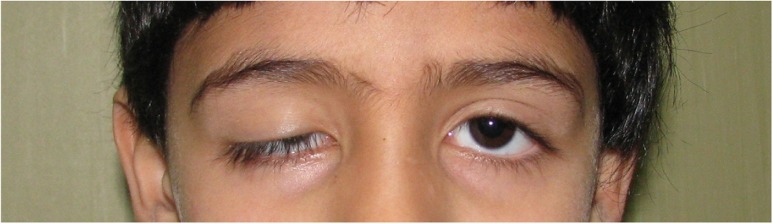
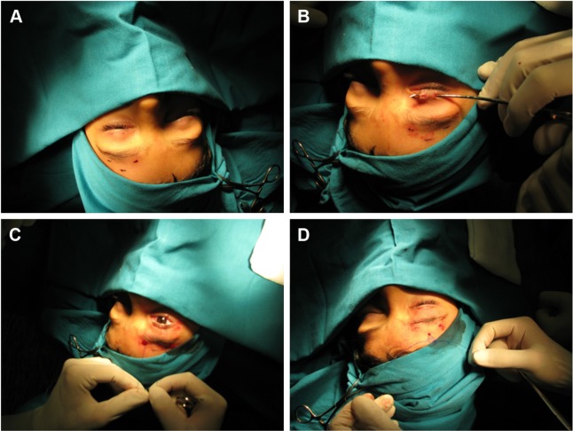
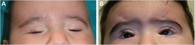
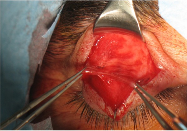
Congenital ptosis is a rare condition characterized by lower positioning of the upper eyelid that is present at birth and is a clinical condition that is persistent if not treated. It may be unilateral or bilateral and may be associated with other ocular disorders or systemic conditions, including Marcus Gunn, Horner, and Duane syndromes. It is a benign condition but causes functional, cosmetic, and psychological problems in children. However, not all patients need to undergo surgery, and usually only patients at risk of amblyopia need a prompt surgical correction, while in other cases, surgery can be postponed. The grade of ptosis, the eyelid function, and the amblyopic risk are the parameters that affect the ophthalmologist's decision on timing of surgery and the surgical technique to be used. In fact, there are several types of surgical techniques to correct a congenital ptosis, although very often more than one is needed to obtain an acceptable result. This paper reviews the causes of congenital ptosis and associated diseases. Particular emphasis is given to surgical management and different procedures available to correct the upper eyelid anomaly and avoid permanent damage to visual function.
Keywords: extraocular muscle development; neurologic dysfunction; ptosis; surgical approach.
Conflict of interest statement
Disclosure The authors report no conflicts of interest in this work.
Figures
References
-
- Sakol PJ, Mannor G, Massaro BM. Congenital and acquired blepharoptosis. Curr Opin Ophthalmol. 1999;10(5):335–339. - PubMed
-
- SooHoo JR, Davies BW, Allard FD, Durairaj VD. Congenital ptosis. Surv Ophthalmol. 2014;59(5):483–492. - PubMed
-
- Vestal KP, Seiff SR, Lahey JM. Congenital ptosis in monozygotic twins. Ophthal Plast Reconstr Surg. 1990;6(4):265–268. - PubMed
-
- Von Noorden GK, Maumenee E. Clinical observations on stimulus-deprivation amblyopia (amblyopia ex anopsia) Am J Ophthalmol. 1968;65(2):220–224. - PubMed
-
- Berry-Brincat A, Willshaw H. Paediatric blepharoptosis: a 10-year review. Eye (Lond) 2009;23(7):1554–1559. - PubMed
Publication types
LinkOut - more resources
Full Text Sources
Other Literature Sources
