. 2017 Feb 23:8:73.
doi: 10.3389/fphar.2017.00073. eCollection 2017.

Embryonic Stem Cell-Derived Neurons Grown on Multi-Electrode Arrays as a Novel In vitro Bioassay for the Detection of Clostridium botulinum Neurotoxins

Affiliations

Embryonic Stem Cell-Derived Neurons Grown on Multi-Electrode Arrays as a Novel In vitro Bioassay for the Detection of Clostridium botulinum Neurotoxins

Stephen P Jenkinson et al. Front Pharmacol. .

Abstract

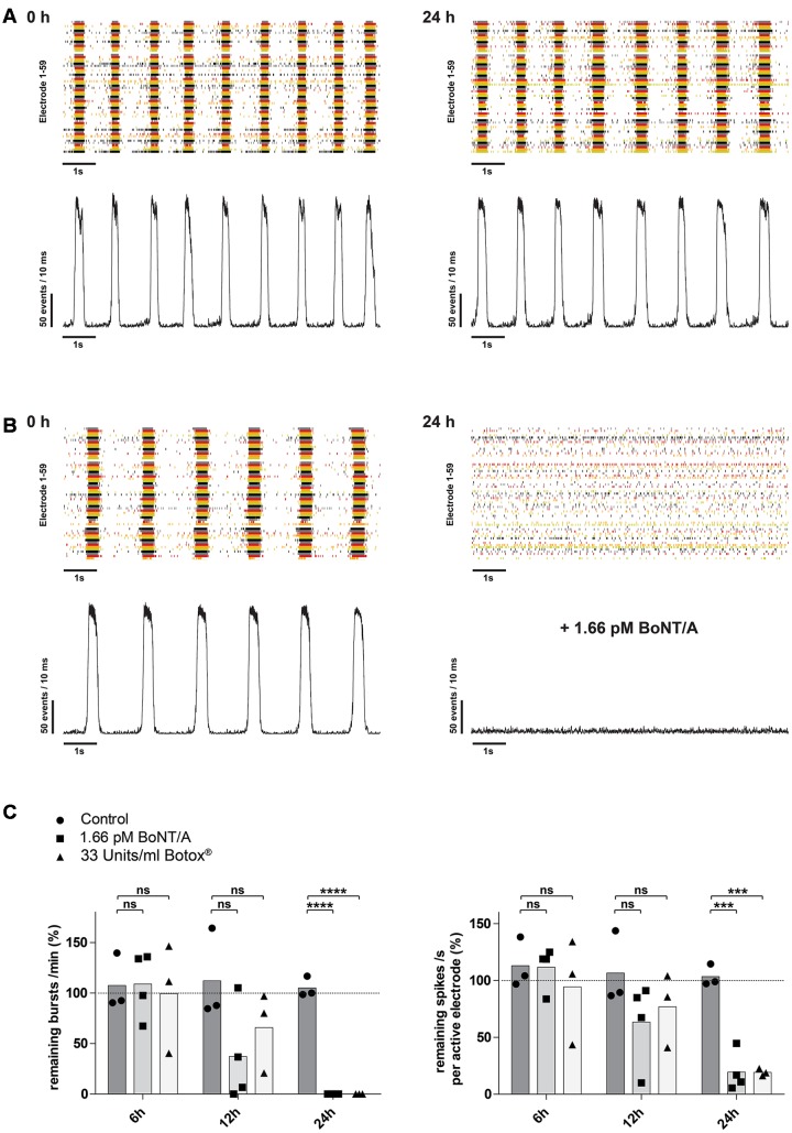
Clostridium botulinum neurotoxins (BoNTs) are the most poisonous naturally occurring protein toxins known to mankind and are the causative agents of the severe and potentially life-threatening disease botulism. They are also known for their application as cosmetics and as unique bio-pharmaceuticals to treat an increasing number of neurological and non-neurological disorders. Currently, the potency of biologically active BoNT for therapeutic use is mainly monitored by the murine LD50-assay, an ethically disputable test causing suffering and death of a considerable number of mice. The aim of this study was to establish an in vitro assay as an alternative to the widely used in vivo mouse bioassay. We report a novel BoNT detection assay using mouse embryonic stem cell-derived neurons (mESN) cultured on multi-electrode arrays (MEAs). After 21 days in culture, the mESN formed a neuronal network showing spontaneous bursting activity based on functional synapses and express the necessary target proteins for BoNTs. Treating cultures for 6 h with 16.6 pM of BoNT serotype A and incubation with 1.66 pM BoNT/A or 33 Units/ml of Botox® for 24 h lead to a significant reduction of both spontaneous network bursts and average spike rate. This data suggests that mESN cultured on MEAs pose a novel, biologically relevant model that can be used to detect and quantify functional BoNT effects, thus accelerating BoNT research while decreasing animal use.

Keywords: BoNT; MEA; botulinum neurotoxins; botulism; embryonic stem cell-derived neurons; in vitro bioassay; multi-electrode array; neuronal network.


Figures

FIGURE 1
FIGURE 1
Representative immunocytochemistry demonstrating the expression of neuronal, synaptic, glutamatergic, GABAergic, and glial markers in cultures 21 days after plating. Neuronal cultures were immunostained against β-III Tubulin (A–F); SNAP-25 (A); SV2 isoform A–C (A,B); polysialo gangliosides GD1a and GT1b/2b (C) Syn1 (D,E); glutamatergic neurons (VGLUT2, D); GABAergic neurons (GAD1, E) and glial cells (GFAP, F). Shown also are DAPI nuclear staining and the merged images. Scale bar is 10 μm (A–E), respectively, 25 μm for the bottom panel (F).
FIGURE 2
FIGURE 2
Electrophysiological characterization of mESN cultured on MEAs. (A, top) Raster plot of 62 single electrodes (represented by different colored lines) showing high spontaneous neuronal activity of a culture 21 days after plating. The formation of bursts is based on spontaneous intrinsic activity and recurrent excitation of the neuronal network through synaptic transmission. Events represent the neuronal activity recognized by the detector (see Materials and Methods). (A, bottom) 10 s excerpt of the raster plot showing individual bursts. (B) Network activity plot. This plot visualizes the amount of activity in the whole neuronal culture network during the time illustrated in the raster plot shown in (A). It shows the number of events detected from all active electrodes with a sliding window of 10 ms shifted by a 1 ms step. Visible by the underlines are the beginning and ending of detected oscillating burst activities. (C) Raw data traces from three single MEA electrodes. (D) Schematic representation of mESN cultured for 21 days on a MEA immunostained for β-III Tubulin (green), GFAP (red), and DAPI (blue). Highlighted in white are the positions of each electrode. Scale bar is 20 μm.
FIGURE 3
FIGURE 3
mESN cultured on MEAs for 21 days show functional glutamatergic and GABAergic synapses with network characteristics. (A) Event raster plot and corresponding network activity plot of activity recorded following the addition of gabazine, strychnine, APV, and CNQX (highlighted by the blue bar). This combination of receptor antagonists leads to an inhibition of synaptic transmission and thus preventing the formation of bursts as well as resulting in a dramatic decrease of the average spike rate. (B) Note that under these conditions asynchronous intrinsic activity is still visible on several electrodes. Raw data traces are shown for three electrodes (E 36, E 39, E 42) as well as for one electrode (E 36) after application of 1 μM TTX. (C) Network activity plot showing the activity prior (top) and after treatment with 10 μM gabazine and 1 μM strychnine. (D) Note the slight increase in burst (left) as well as average spike rate (right). Data are given as mean; paired t-test, two tailed; ∗∗P < 0.01; ns signifies as not significant.
FIGURE 4
FIGURE 4
mESN grown on MEAs are susceptible to intoxication by BoNT/A in a dose-dependent manner. Treatment and incubation for 6 h with 16.6 and 166 pM BoNT/A resulted in a significant decrease of the burst rate and the mean spike rate. Cultures receiving no toxin served as control. (A) Reduction of the burst rate compared to the control group. (B) Reduction of the mean spike rate compared to the control group. Mann–Whitney two tailed test; P < 0.05; ∗∗P < 0.01; ∗∗∗P < 0.001; ns signifies as not significant. (C) Treatment and incubation with 166 pM heat inactivated BoNT/A showed no significant difference in burst (left) as well as average spike rate (right). Paired t-test, two tailed; ns signifies as not significant.
FIGURE 5
FIGURE 5
Time-dependent reduction of synaptic transmission upon addition of BoNT/A holotoxin and complex. (A) Raster plot and corresponding network activity plot showing spontaneous burst activity of a 21-day-old control culture prior (left) and 24 h after receiving 10 μl of PBS (right). (B) Upon treatment and incubation with 1.66 pM BoNT/A for 24 h a complete loss of synchronous burst activity is visible (right). Seen on the left is the same culture prior to addition of the toxin serving as the base activity at 0 h (left). (C) Decrease of the burst rate (left) and the mean spike rate (right) upon treatment and incubation with 1.66 pM BoNT/A or 33 Units/ml Botox® at the indicated time points. Each culture was normalized toward its base activity measured at time point 0 h (highlighted by the dotted line). The reduction of the activity was then compared toward untreated cultures serving as a control. Unpaired t-test, two tailed; ∗∗∗P < 0.001; ∗∗∗∗P < 0.0001; ns signifies as not significant.

Similar articles

Cited by

References

    1. Abranches E., Silva M., Pradier L., Schulz H., Hummel O., Henrique D., et al. (2009). Neural differentiation of embryonic stem cells in vitro: a road map to neurogenesis in the embryo. PLoS ONE 4:e6286 10.1371/journal.pone.0006286 - DOI - PMC - PubMed
    1. Adler S., Bicker G., Bigalke H., Bishop C., Blumel J., Dressler D., et al. (2010). The current scientific and legal status of alternative methods to the LD50 test for botulinum neurotoxin potency testing. The report and recommendations of a ZEBET Expert Meeting. Altern. Lab. Anim. 38 315–330. - PubMed
    1. Akaike N., Ito Y., Shin M. C., Nonaka K., Torii Y., Harakawa T., et al. (2010). Effects of A2 type botulinum toxin on spontaneous miniature and evoked transmitter release from the rat spinal excitatory and inhibitory synapses. Toxicon 56 1315–1326. 10.1016/j.toxicon.2010.07.015 - DOI - PubMed
    1. Arnon S. S., Schechter R., Inglesby T. V., Henderson D. A., Bartlett J. G., Ascher M. S., et al. (2001). Botulinum toxin as a biological weapon: medical and public health management. JAMA 285 1059–1070. - PubMed
    1. Ban J., Bonifazi P., Pinato G., Broccard F. D., Studer L., Torre V., et al. (2007). Embryonic stem cell-derived neurons form functional networks in vitro. Stem Cells 25 738–749. 10.1634/stemcells.2006-0246 - DOI - PubMed