. 2017 Feb 23:8:262.
doi: 10.3389/fmicb.2017.00262. eCollection 2017.

Toll-Like Receptor 2 Is Required for Inflammatory Process Development during Leishmania infantum Infection

Affiliations

Toll-Like Receptor 2 Is Required for Inflammatory Process Development during Leishmania infantum Infection

Laís A Sacramento et al. Front Microbiol. .

Abstract

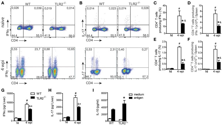
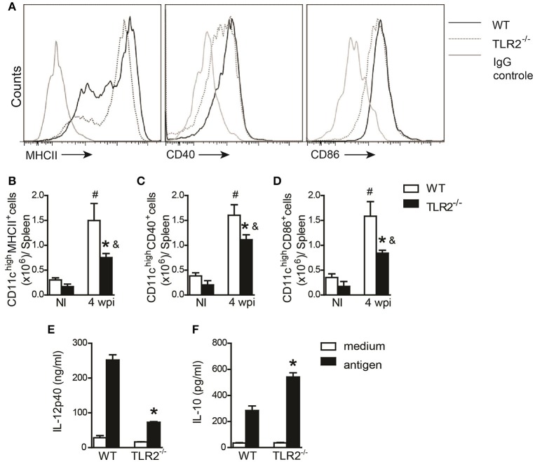
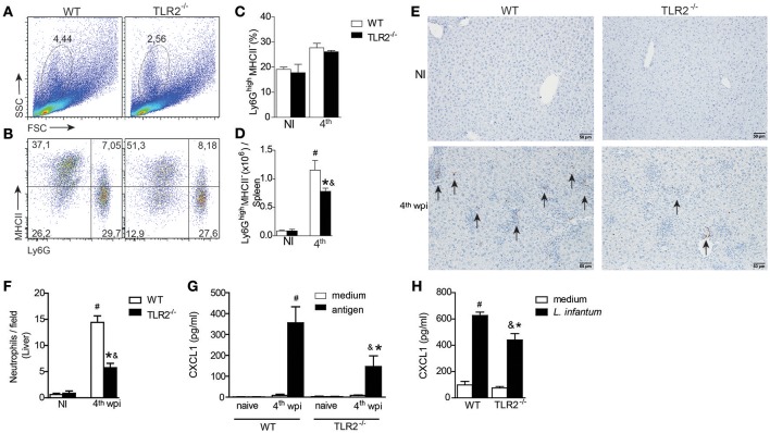
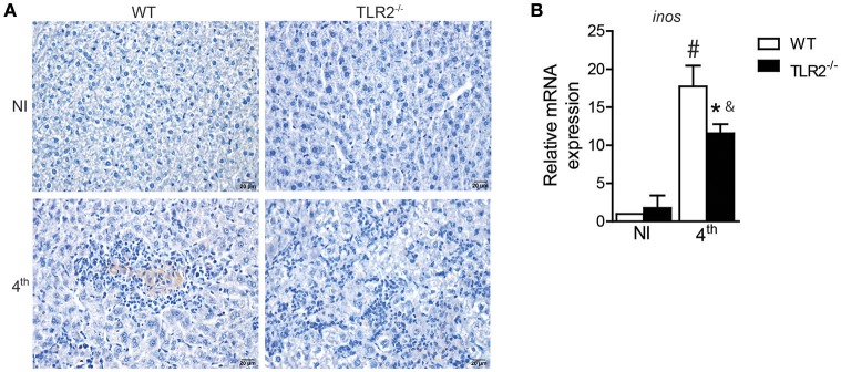
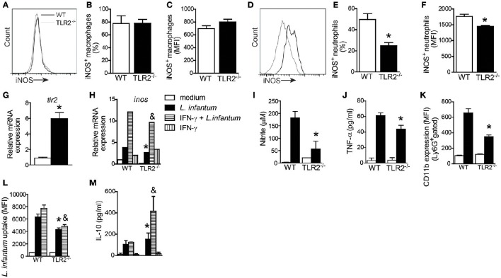
Visceral leishmaniasis (VL) is a chronic and fatal disease caused by Leishmania infantum in Brazil. Leukocyte recruitment to infected tissue is a crucial event for the control of infections such as VL. Among inflammatory cells, neutrophils are recruited to the site of Leishmania infection, and these cells may control parasite replication through oxidative or non-oxidative mechanisms. The recruitment, activation and functions of the neutrophils are coordinated by pro-inflammatory cytokines and chemokines during recognition of the parasite by pattern recognition receptors (PRRs). Here, we demonstrated that the Toll-like receptor 2 (TLR2) signaling pathway contributes to the development of the innate immune response during L. infantum infection. The protective mechanism is related to the appropriate recruitment of neutrophils to the inflammatory site. Neutrophil migration is coordinated by DCs that produce CXCL1 and provide a prototypal Th1 and Th17 environment when activated via TLR2. Furthermore, infected TLR2-/- mice failed to induce nitric oxide synthase (iNOS) expression in neutrophils but not in macrophages. In vitro, infected TLR2-/- neutrophils presented deficient iNOS expression, nitric oxide (NO) and TNF-α production, decreased expression of CD11b and reduced L. infantum uptake capacity. The non-responsive state of neutrophils is associated with increased amounts of IL-10. Taken together, these data clarify new mechanisms by which TLR2 functions in promoting the development of the adaptive immune response and effector mechanisms of neutrophils during L. infantum infection.

Keywords: Leishmania infantum; TLR2; dendritic cell maturation; neutrophil activation; visceral leishmaniasis.


Figures

Figure 1
Figure 1
TLR2 is important for host protection against L. infantum. TLR2 expression in in vitro L. infantum-infected WT BMDCs (1:5) during 24 h (A). The percentage and MFI of DC (CD11chigh) expression of TLR2 was determined by flow cytometry (B). tlr2 mRNA expression in the spleens and livers from WT mice at the 4th wpi was evaluated by real-time PCR and normalized to the constitutively expressed HPRT and GAPDH genes, respectively (C). Parasite burdens in the spleen and liver were determined in WT and TLR2−/− mice at the 4th and 6th weeks p.i. by limiting dilution (D). Representative photomicrography of stained liver sections is shown at 20 times the original magnification (E). The inflammation areas in WT (open circles) and TLR2−/− (closed circles) mice were quantified using Leica Qwin software and are shown as the frequency distribution of the lesion areas (F). Data are expressed as the mean ± SEM and are representative of three independent experiments. *P < 0.05 [Student's t-test (B,C,F) or two-way ANOVA with Tukey's post-hoc test (D)] is relative to the control group.
Figure 2
Figure 2
TLR2 participates in Th1 and Th17 immune response development during L. infantum infection. The spleen cells from uninfected (NI) or 4 wpi WT and TLR2−/− mice were restimulated in vitro with PMA (50 ng/mL) and ionomycin (500 ng/mL) for 4 h and analyzed for intracellular cytokine production by flow cytometry. Dot plots represent the frequency of naïve (above) and infected (below) CD4+ T cell-producing IFN-γ (A) and IL-17 (B), and the graph bars represent the percentage (C,E) and the absolute number (D,F) of these cells. The liver fragments from the WT or TLR2−/− mice at the 4th wpi were collected and weighed for the determination of IFN-γ (G) and IL-17 (H) by ELISA in the homogenate supernatants. In vitro restimulation of spleen cells with the L. infantum antigen (50 μg/mL) or medium for 72 h, and the levels of IL-10 (I) were measured in the culture supernatants by an ELISA. Data are expressed as the mean ± SEM and are representative of three independent experiments, N = 4–5. *P < 0.05 compared to infected WT (A–G) or compared to antigen-stimulated WT (H), #P < 0.05 compared to uninfected WT, &P < 0.05 compared to uninfected TLR2−/− (two-way ANOVA with Tukey's post-hoc test).
Figure 3
Figure 3
DC activation during L. infantum infection is dependent on TLR2. In vivo surface markers of DCs from uninfected (NI) and at 4th wpi WT and TLR2−/− mice were determined by flow cytometry. Histograms demonstrate the costimulatory molecules in the CD11chigh population (A) and bar graphs represent the percentage of MHCII (B), CD40 (C), and CD86 (D). All analyses were performed on CD11b+CD11chigh gated cells. WT and TLR2−/− BMDCs were infected with L. infantum (5:1) or medium for 24 h and IL-12p40 (E) and IL-10 (F) levels in the supernatants were measured by ELISA. Data are expressed as the mean ± SEM and are representative of three independent experiments, N = 4–5. *P < 0.05 compared to infected WT (B–D) or compared to stimulated WT (E,F), #P < 0.05 compared to uninfected WT, &P < 0.05 compared to uninfected TLR2−/− (two-way ANOVA with Tukey's post-hoc test).
Figure 4
Figure 4
TLR2 induces neutrophil migration to target organs of L. infantum infection. The neutrophil population was gated based on their characteristic size (FSC) and granularity (SSC) (A). The dot plots represent the frequency of neutrophil population characterized by Ly6G+MHCII by flow cytometry (B). The bar graphs represent the percentage (C) and absolute number (D) of the Ly6G+MHCII population in the spleens from WT and TLR2−/− mice at the 4th wpi or from uninfected mice (NI). Neutrophil numbers in liver sections of WT and TLR2−/− mice at the 4th wpi or from uninfected mice (NI) by immunohistochemistry with anti-mouse 7/4 antibody arrows indicate 7/4+ cells (E). Quantification of 7/4+ cells present in the liver tissue of WT and TLR2−/− mice at the 4th wpi or from NI mice (F). Arrays show the 7/4+ cells. CXCL1 measured from the supernatant of in vitro-restimulated spleen cells with the L. infantum antigen (50 μg/mL) or medium for 72 h (G) or from L. infantum in vitro-infected DCs (H) by ELISA. Data are expressed as the mean ± SEM and are representative of three (A–D) and two (E,F) independent experiments, N = 4–5. *P < 0.05 compared to infected WT (D,F) or compared to stimulated WT (G), #P < 0.05 compared to uninfected WT, &P < 0.05 compared to uninfected TLR2−/− (two-way ANOVA with Tukey's post-hoc test).
Figure 5
Figure 5
iNOS expression in target organs of L. infantum infection is dependent on TLR2. iNOS staining in liver sections of WT and TLR2−/− mice at the 4th wpi or from uninfected mice (NI) by immunohistochemistry with anti-mouse iNOS antibody (A). inos mRNA expression in the spleen from WT mice at the 4th wpi was evaluated by real-time PCR and normalized to the constitutively expressed HPRT (B). Data are expressed as the mean ± SEM and are representative of three (A) and two (B) independent experiments, N = 4–5. *P < 0.05 compared to infected WT, #P < 0.05 compared to uninfected WT, &P < 0.05 compared to uninfected TLR2−/− (two-way ANOVA with Tukey's post-hoc test).
Figure 6
Figure 6
TLR2 promotes nitric oxide and TNF production by neutrophils and killing of L. infantum. Spleen cells of WT and TLR2−/− mice were isolated from infected mice and stained for iNOS in CD11cMHCII+CD11b+ cell (macrophage) and Ly6G+MHCII cell (neutrophil) populations. Representative histograms demonstrate the iNOS expression gated in CD11cMHCII+CD11b+ cell (A) and Ly6G+MHCII cell (D) populations. The bar graphs represent the percentage and MFI of iNOS+ macrophages (B,C) and iNOS+ neutrophils (E,F). Neutrophils were isolated from bone marrow and infected with promastigote forms of L. infantum (5:1) or uninfected (medium). After 4 h, tlr2 (G) and inos (H) mRNA expression was analyzed by quantitative PCR, and the supernatant was collected for nitric oxide (I) and TNF (J) production, or the cells were harvested for CD11b to evaluate the neutrophil activation by flow cytometry (K). For the L. infantum uptake assay, the neutrophils were previously primed with IFN-γ (100 ng/mL) for 1 h and cultured with CFSE stained L. infantum promastigote forms (5:1) for 4 h (L) and IL-10 from the supernatant was measured (M). Data are expressed as the mean ± SEM. *P < 0.05 compared to infected WT (E,F) or WT stimulated with L. infantum (G–M), &P < 0.05 compared to WT stimulated with IFN-γ + L. infantum (H,L,M) [Student's t-test (E–G) or two-way ANOVA with Tukey's post-hoc test (H–M)] relative to the control group.

Similar articles

Cited by

References

    1. Akira S., Uematsu S., Takeuchi O. (2006). Pathogen recognition and innate immunity. Cell 124, 783–802. 10.1016/j.cell.2006.02.015 - DOI - PubMed
    1. Alexander J., Bryson K. (2005). T helper (h) 1/Th2 and Leishmania: paradox rather than paradigm. Immunol. Lett. 99, 17–23. 10.1016/j.imlet.2005.01.009 - DOI - PubMed
    1. Applequist S. E., Wallin R. P., Ljunggren H. G. (2002). Variable expression of Toll-like receptor in murine innate and adaptive immune cell lines. Int. Immunol. 14, 1065–1074. 10.1093/intimm/dxf069 - DOI - PubMed
    1. Ashour D. S. (2015). Toll-like receptor signaling in parasitic infections. Expert Rev. Clin. Immunol. 11, 771–780. 10.1586/1744666X.2015.1037286 - DOI - PubMed
    1. Assis R. R., Ibraim I. C., Noronha F. S., Turco S. J., Soares R. P. (2012). Glycoinositolphospholipids from Leishmania braziliensis and L. infantum: modulation of innate immune system and variations in carbohydrate structure. PLoS Negl. Trop. Dis. 6:e1543. 10.1371/journal.pntd.0001543 - DOI - PMC - PubMed