T-Cells Specific for a Self-Peptide of ApoB-100 Exacerbate Aortic Atheroma in Murine Atherosclerosis
- PMID: 28280493
- PMCID: PMC5322236
- DOI: 10.3389/fimmu.2017.00095
T-Cells Specific for a Self-Peptide of ApoB-100 Exacerbate Aortic Atheroma in Murine Atherosclerosis
Abstract
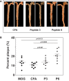
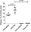
On the basis of mouse I-Ab-binding motifs, two sequences of the murine apolipoprotein B-100 (mApoB-100), mApoB-1003501-3515 (designated P3) and mApoB-100978-992 (designated P6), were found to be immunogenic. In this report, we show that P6 is also atherogenic. Immunization of Apoe-/- mice fed a high-fat diet (HFD) with P6 resulted in enhanced development of aortic atheroma as compared to control mice immunized with an irrelevant peptide MOG35-55 or with complete Freund's adjuvant alone. Adoptive transfer of lymph node cells from P6-immunized donor mice to recipients fed an HFD caused exacerbated aortic atheromas, correlating P6-primed cells with disease development. Finally, P6-specific T cell clones were generated and adoptive transfer of T cell clones into recipients fed an HFD led to significant increase in aortic plaque coverage when compared to control animals receiving a MOG35-55-specific T cell line. Recipient mice not fed an HFD, however, did not exhibit such enhancement, indicating that an inflammatory environment facilitated the atherogenic activity of P6-specific T cells. That P6 is identical to or cross-reacts with a naturally processed peptide of ApoB-100 is evidenced by the ability of P6 to stimulate the proliferation of T cells in the lymph node of mice primed by full-length human ApoB-100. By identifying an atherogenic T cell epitope of ApoB-100 and establishing specific T cell clones, our studies open up new and hitherto unavailable avenues to study the nature of atherogenic T cells and their functions in the atherosclerotic disease process.
Keywords: T-cell; adoptive transfer; atherosclerosis; clones; epitopes; peptides.
Figures


Similar articles
-
A clinically applicable adjuvant for an atherosclerosis vaccine in mice.Eur J Immunol. 2018 Sep;48(9):1580-1587. doi: 10.1002/eji.201847584. Epub 2018 Aug 12. Eur J Immunol. 2018. PMID: 29932463 Free PMC article.
-
Identification of apoB-100 Peptide-Specific CD8+ T Cells in Atherosclerosis.J Am Heart Assoc. 2017 Jul 15;6(7):e005318. doi: 10.1161/JAHA.116.005318. J Am Heart Assoc. 2017. PMID: 28711866 Free PMC article.
-
Role of an immunodominant T cell epitope of the P6 protein of nontypeable Haemophilus influenzae in murine protective immunity.Vaccine. 2005 May 20;23(27):3590-6. doi: 10.1016/j.vaccine.2005.01.151. Vaccine. 2005. PMID: 15855018
-
Single-cell transcriptomes and T cell receptors of vaccine-expanded apolipoprotein B-specific T cells.Front Cardiovasc Med. 2023 Jan 5;9:1076808. doi: 10.3389/fcvm.2022.1076808. eCollection 2022. Front Cardiovasc Med. 2023. PMID: 36684560 Free PMC article.
-
Lipoprotein size and atherosclerosis susceptibility in Apoe(-/-) and Ldlr(-/-) mice.Arterioscler Thromb Vasc Biol. 2001 Oct;21(10):1567-70. doi: 10.1161/hq1001.097780. Arterioscler Thromb Vasc Biol. 2001. PMID: 11597927 Review.
Cited by
-
Atherosclerosis: from lipid-lowering and anti-inflammatory therapies to targeting arterial retention of ApoB-containing lipoproteins.Front Immunol. 2025 Jun 9;16:1485801. doi: 10.3389/fimmu.2025.1485801. eCollection 2025. Front Immunol. 2025. PMID: 40552286 Free PMC article. Review.
-
ApoB-Specific CD4+ T Cells in Mouse and Human Atherosclerosis.Cells. 2021 Feb 19;10(2):446. doi: 10.3390/cells10020446. Cells. 2021. PMID: 33669769 Free PMC article. Review.
-
A clinically applicable adjuvant for an atherosclerosis vaccine in mice.Eur J Immunol. 2018 Sep;48(9):1580-1587. doi: 10.1002/eji.201847584. Epub 2018 Aug 12. Eur J Immunol. 2018. PMID: 29932463 Free PMC article.
-
Regulatory CD4+ T Cells Recognize Major Histocompatibility Complex Class II Molecule-Restricted Peptide Epitopes of Apolipoprotein B.Circulation. 2018 Sep 11;138(11):1130-1143. doi: 10.1161/CIRCULATIONAHA.117.031420. Circulation. 2018. PMID: 29588316 Free PMC article.
-
[Immunization as Treatment for arteriosclerosis-a realistic vision?].Herz. 2019 Apr;44(2):93-95. doi: 10.1007/s00059-019-4793-8. Herz. 2019. PMID: 30923854 German. No abstract available.
References
Grants and funding
LinkOut - more resources
Full Text Sources
Other Literature Sources
Research Materials
Miscellaneous

