Myeloid and T Cell-Derived TNF Protects against Central Nervous System Tuberculosis
- PMID: 28280495
- PMCID: PMC5322283
- DOI: 10.3389/fimmu.2017.00180
Myeloid and T Cell-Derived TNF Protects against Central Nervous System Tuberculosis
Abstract
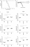
Tuberculosis of the central nervous system (CNS-TB) is a devastating complication of tuberculosis, and tumor necrosis factor (TNF) is crucial for innate immunity and controlling the infection. TNF is produced by many cell types upon activation, in particularly the myeloid and T cells during neuroinflammation. Here we used mice with TNF ablation targeted to myeloid and T cell (MT-TNF-/-) to assess the contribution of myeloid and T cell-derived TNF in immune responses during CNS-TB. These mice exhibited impaired innate immunity and high susceptibility to cerebral Mycobacterium tuberculosis infection, a similar phenotype to complete TNF-deficient mice. Further, MT-TNF-/- mice were not able to control T cell responses and cytokine/chemokine production. Thus, our data suggested that collective TNF production by both myeloid and T cells are required to provide overall protective immunity against CNS-TB infection.
Keywords: CNS; Mycobacterium tuberculosis; T cell; myeloid; tumor necrosis factor.
Figures


Similar articles
-
TNF-dependent regulation and activation of innate immune cells are essential for host protection against cerebral tuberculosis.J Neuroinflammation. 2015 Jun 26;12:125. doi: 10.1186/s12974-015-0345-1. J Neuroinflammation. 2015. PMID: 26112704 Free PMC article.
-
[Frontier of mycobacterium research--host vs. mycobacterium].Kekkaku. 2005 Sep;80(9):613-29. Kekkaku. 2005. PMID: 16245793 Japanese.
-
Myd88 Initiates Early Innate Immune Responses and Promotes CD4 T Cells during Coronavirus Encephalomyelitis.J Virol. 2015 Sep;89(18):9299-312. doi: 10.1128/JVI.01199-15. Epub 2015 Jul 1. J Virol. 2015. PMID: 26136579 Free PMC article.
-
[Up-to-date understanding of tuberculosis immunity].Kekkaku. 2003 Jan;78(1):51-5. Kekkaku. 2003. PMID: 12683337 Review. Japanese.
-
[Immunity against Mycobacterium tuberculosis (introduction)].Kekkaku. 2010 Jun;85(6):501-8. Kekkaku. 2010. PMID: 20662245 Review. Japanese.
Cited by
-
Immune control of Mycobacterium tuberculosis is dependent on both soluble TNFRp55 and soluble TNFRp75.Immunology. 2021 Nov;164(3):524-540. doi: 10.1111/imm.13385. Epub 2021 Jul 8. Immunology. 2021. PMID: 34129695 Free PMC article.
-
Mycobacterium Tuberculosis and Interactions with the Host Immune System: Opportunities for Nanoparticle Based Immunotherapeutics and Vaccines.Pharm Res. 2018 Nov 8;36(1):8. doi: 10.1007/s11095-018-2528-9. Pharm Res. 2018. PMID: 30411187 Free PMC article. Review.
-
Neutrophil-Mediated Immunopathology and Matrix Metalloproteinases in Central Nervous System - Tuberculosis.Front Immunol. 2022 Jan 12;12:788976. doi: 10.3389/fimmu.2021.788976. eCollection 2021. Front Immunol. 2022. PMID: 35095865 Free PMC article. Review.
-
Complete ablation of tumor necrosis factor decreases the production of IgA, IgG, and IgM in experimental central nervous system tuberculosis.Iran J Basic Med Sci. 2020 May;23(5):680-690. doi: 10.22038/ijbms.2020.37947.9021. Iran J Basic Med Sci. 2020. PMID: 32742607 Free PMC article.
-
Bioinformation Analysis Reveals IFIT1 as Potential Biomarkers in Central Nervous System Tuberculosis.Infect Drug Resist. 2022 Jan 6;15:35-45. doi: 10.2147/IDR.S328197. eCollection 2022. Infect Drug Resist. 2022. PMID: 35027832 Free PMC article.
References
-
- Lin PL, Myers A, Smith L, Bigbee C, Bigbee M, Fuhrman C, et al. Tumor necrosis factor neutralization results in disseminated disease in acute and latent Mycobacterium tuberculosis infection with normal granuloma structure in a cynomolgus macaque model. Arthritis Rheum (2010) 62:340–50.10.1002/art.27271 - DOI - PMC - PubMed
LinkOut - more resources
Full Text Sources
Other Literature Sources

