. 2017:2017:4123854.
doi: 10.1155/2017/4123854. Epub 2017 Feb 9.

Reactive Oxygen Species Mediated Prostaglandin E2 Contributes to Acute Response of Epithelial Injury

Affiliations

Reactive Oxygen Species Mediated Prostaglandin E2 Contributes to Acute Response of Epithelial Injury

Yi-Ping Hu et al. Oxid Med Cell Longev. 2017.

Abstract

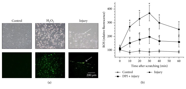
Reactive oxygen species (ROS) generated after tissue injury play a crucial role during wound healing through initiating acute inflammation, clarifying infection and dead tissue, and mediating various intracellular signal transduction. Prostaglandin E2 (PGE2) has been identified as one of the major factors responsible for inflammation and tissue repair. In this study, we tested our hypothesis that ROS produced by damaged human keratinocytes induces the synthesis of PGE2. In vitro epithelial wounding model was used to observe the production of ROS and secretion of PGE2 as well as the involved signal pathway. The mechanical injury caused the rapid production of ROS in in vitro cultured keratinocytes, which was significantly blocked by an inhibitor of nicotinamide adenine dinucleotide phosphate oxidase. The increased intracellular ROS caused by mechanical injury stimulates PGE2 production in a time-dependent manner via the activation of cyclooxygenase-2 (COX-2), which was stimulated by phosphorylation of extracellular signal-regulated protein kinase (ERK). These results indicate ROS-induced ERK activation leading to the activation of COX-2 and the synthesis of PGE2 in human keratinocytes responding to mechanical injury in the acute phase.


Conflict of interest statement

The authors declare that there is no conflict of interests regarding the publication of this paper.

Figures

Figure 1
Figure 1
Rapid formation of ROS induced by scratching injury in HaCat cells. (a) The intracellular ROS were visualized by the reaction with probe carboxy-H2DCFDA under fluorescent microscopy. Cells without damage worked as negative control, while H2O2 treated cells were used as positive control. (b) The quantities of ROS were shown after scratching for undamaged control cells, damaged cells, and damaged cells plus DPI treatment. Data are representative of three independent experiments with triplicate samples. and # indicate p < 0.05 compared with undamaged control cells and with scratched cells, respectively.
Figure 2
Figure 2
Scratching-injury-induced PGE2 production and the despondence on ROS. (a) PGE2 secretion by HaCat cells after scratches. PGE2 was measured by ELISA in different periods of time as indicated. (b) Suppressed secretion PGE2 by NOX/ROS inhibitor. Values are means ± SEMs of three replicates. and # indicate p < 0.05 compared with undamaged control cells and with scratched cells, respectively.
Figure 3
Figure 3
Participation of ERK activation in scratching injury-induced release of ROS and PGE2. (a) The change of p-ERK and ERK in HaCat cells treated with scratching injury for different periods of time as indicated. The protein levels of p-ERK and ERK were determined by immunoblot analysis and normalized to β-tubulin. (b) The level of p-ERK was decreased in HaCat cells preincubated with different concentrations of DPI for 30 min followed by scratches for 0.5 hr. (c) The dependence of PGE2 production caused by scratching injury on ERK activation. Values are means ± SEMs of three replicates. and # indicate p < 0.05 compared with undamaged control cells and with scratched cells, respectively.
Figure 4
Figure 4
Injury-induced PGE2 release mediated by COX-2, not COX-1. (a) COX-1 and COX-2 activity of uninjured or injured HaCat cells were measured by COX activity assay. (b) COX-2 expression was increased in HaCat cells treated with scratching injury for different periods of time as indicated. The protein levels of COX-2 were determined by immunoblot analysis and normalized to β-tubulin. (c) The induced expression of COX-2 in HaCat cells was suppressed by NOX/ROS inhibitor DPI. (d) The induced expression of COX-2 in HaCat cells was suppressed by ERK inhibitor. (e) The induced expression of COX-2 in HaCat cells was suppressed by a COX-2 inhibitor. Values are means ± SEMs of three replicates. and # indicate p < 0.05 compared with undamaged control cells and with scratched cells, respectively.

References

    1. Niethammer P., Grabher C., Look A. T., Mitchison T. J. A tissue-scale gradient of hydrogen peroxide mediates rapid wound detection in zebrafish. Nature. 2009;459(7249):996–999. doi: 10.1038/nature08119. - DOI - PMC - PubMed
    1. Roy S., Khanna S., Nallu K., Hunt T. K., Sen C. K. Dermal wound healing is subject to redox control. Molecular Therapy. 2006;13(1):211–220. doi: 10.1016/j.ymthe.2005.07.684. - DOI - PMC - PubMed
    1. Oktyabrsky O. N., Smirnova G. V. Redox regulation of cellular functions. Biochemistry (Moscow) 2007;72(2):132–145. doi: 10.1134/S0006297907020022. - DOI - PubMed
    1. Fu X.-J., Peng Y.-B., Hu Y.-P., Shi Y.-Z., Yao M., Zhang X. NADPH oxidase 1 and its derived reactive oxygen species mediated tissue injury and repair. Oxidative Medicine and Cellular Longevity. 2014;2014:10. doi: 10.1155/2014/282854.282854 - DOI - PMC - PubMed
    1. Jiang F., Zhang Y., Dusting G. J. NADPH oxidase-mediated redox signaling: roles in cellular stress response, stress tolerance, and tissue repair. Pharmacological Reviews. 2011;63(1):218–242. doi: 10.1124/pr.110.002980. - DOI - PubMed

LinkOut - more resources