. 2016 Dec;1(7):606-616.
doi: 10.1016/j.jacbts.2016.07.009. Epub 2016 Nov 16.

Inhibition of Interleukin 17-A but not Interleukin-17F Signaling Lowers Blood Pressure and Reduces End-organ Inflammation in Angiotensin II-induced Hypertension

Affiliations

Inhibition of Interleukin 17-A but not Interleukin-17F Signaling Lowers Blood Pressure and Reduces End-organ Inflammation in Angiotensin II-induced Hypertension

Mohamed A Saleh et al. JACC Basic Transl Sci. 2016 Dec.

Abstract

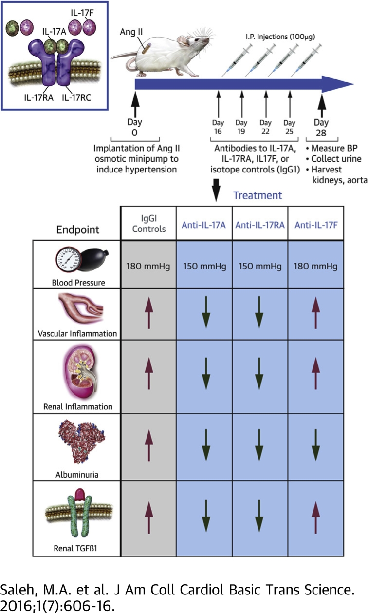
Objectives: To characterize the T cell subsets producing interleukin 17 (IL-17) isoforms A and F in hypertensive kidneys and vessels and determine whether inhibition of IL-17 signaling lowers blood pressure and end-organ damage in a mouse model of hypertension.

Background: T cell derived cytokines play a central role in the pathophysiology of hypertension and contribute to end-organ dysfunction. We previously showed that mice genetically deficient in IL-17A exhibited blunted hypertension and reduced renal and vascular dysfunction in response to angiotensin II (Ang II) infusion. Monoclonal antibodies to IL-17 isoforms or the IL-17 receptor are emerging as novel therapeutics for psoriasis and related autoimmune disorders.

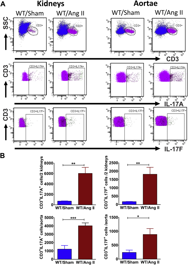
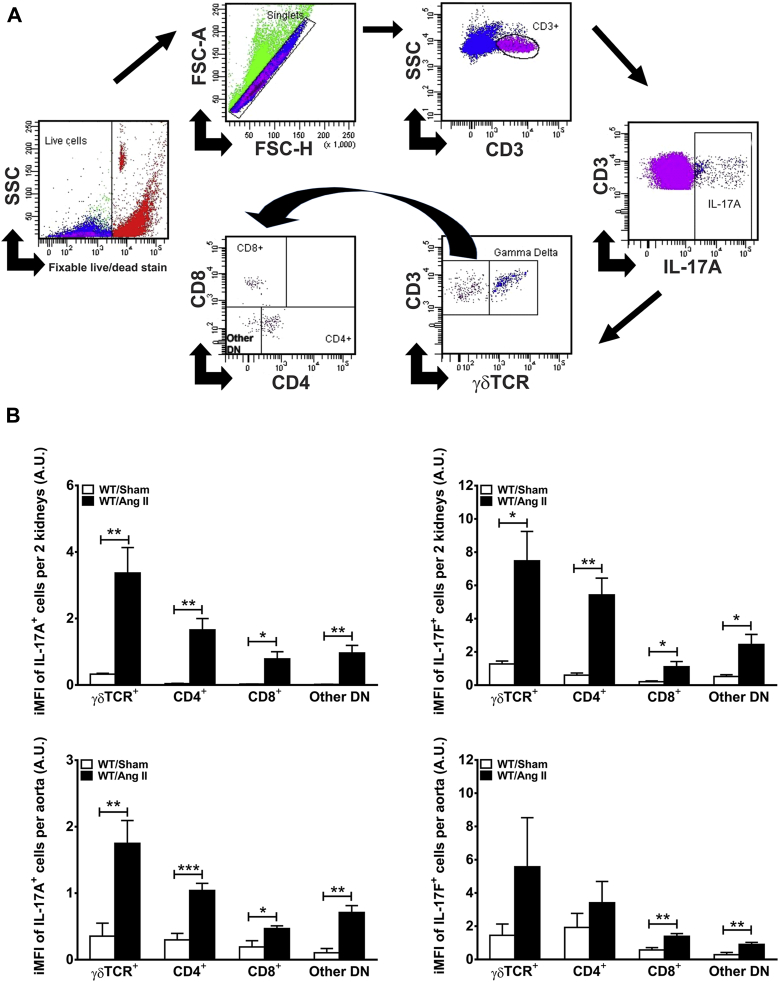
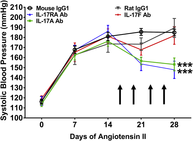
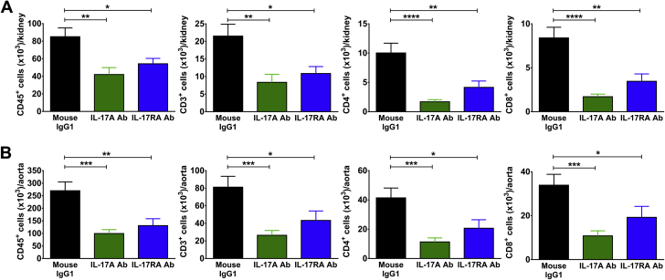
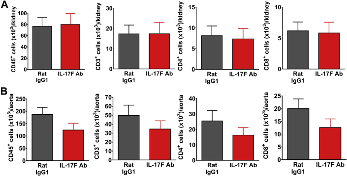
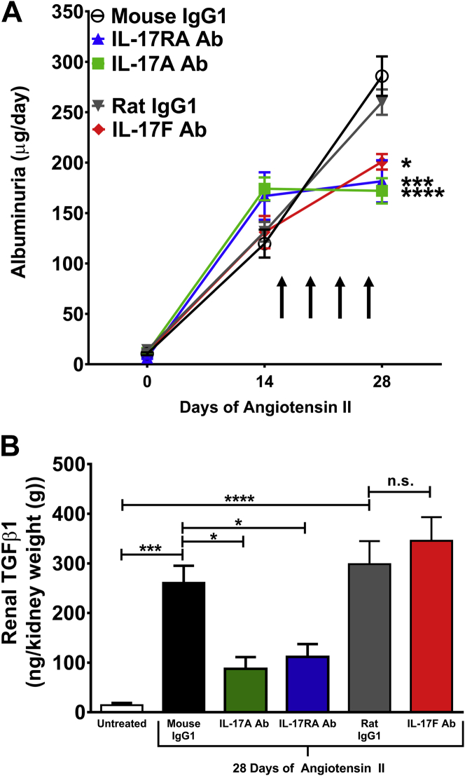
Methods and results: Mice were infused with Ang II for 4 weeks to induce hypertension. Using flow cytometry and intracellular staining, we determined that the primary T cell subsets producing IL-17A and IL-17F in the kidney and aorta are gamma delta (γδ) T cells and CD4+ T helper 17 (TH17) cells. Monoclonal antibodies were administered twice weekly starting 2 weeks after the onset of Ang II infusion. Antibodies to IL-17A or the IL-17 receptor A subunit (IL-17RA), but not IL-17F, lowered blood pressure by 30 mmHg, attenuated renal and vascular lymphocyte infiltration, and reduced renal transforming growth factor beta (TGFβ) levels (a marker of renal fibrosis) compared to control IgG1 antibodies. Inhibition of IL-17 signaling also blunted the progression of albuminuria.

Conclusions: Monoclonal antibodies to IL-17A or IL-17RA, but not IL-17F, may be a useful adjunct treatment for hypertension and the associated end-organ dysfunction.

Keywords: T lymphocytes; angiotensin II; gamma delta T cells; hypertension; interleukin 17A; interleukin 17F; interleukin 17RA receptor.


Figures

None
Graphical abstract
Figure 1
Figure 1
Angiotensin II Increases IL-17A– and IL-17F–Producing T Cells in Mouse Kidneys and Aortae (A) Representative flow cytometry analysis of renal and aortic T cells isolated from mice after 28 days of vehicle (Sham) or angiotensin II (Ang II) infusion. (B) Quantification of the total number of interleukin (IL)-17A– or IL-17F–producing cells in kidneys and aortae (n = 5 to 6 per group). Data were analyzed using Student t test and expressed as mean ± SEM. *p < 0.05; **p < 0.01; ***p < 0.001 vs. WT/Sham. WT = wild type.
Figure 2
Figure 2
γδ T Cells and TH17 Cells are the Major Sources of IL-17A and IL-17F in the Kidney and Vasculature Following Ang II Infusion (A) Representative example of flow cytometry gating strategy for the quantification of IL-17A–producing CD3+ T cells in the kidney of an Ang II–treated mouse. A similar strategy was used for IL-17F and for aortic samples. (B) Quantification of the integrated mean fluorescence intensity (iMFI) of IL-17A– or IL-17F–producing subsets of T cells (γδ T cell receptor [TCR]+, CD4+, CD8+, and other double-negative T cells [other DN]). Data were analyzed using Student t test and expressed in arbitrary units (A.U.) as mean ± SEM (n = 5 to 6 per group). *p < 0.05; **p < 0.01; ***p < 0.001 versus WT/Sham. Abbreviations as in Figure 1.
Figure 3
Figure 3
Monoclonal Antibodies to IL-17A or the IL-17RA Receptor Subunit, But Not IL-17F, Lowers Blood Pressure in Response to Ang II Infusion Systolic blood pressure was measured at baseline and weekly during 28 days of Ang II infusion. Arrows indicate timing of antibody administration. Data are expressed as mean ± SEM and were analyzed at day 28 using 1-way analysis of variance followed by Holm-Sidak post hoc test (n = 7 to 20 per group). ***p < 0.001 vs. mouse IgG1. Ab = antibody; other abbreviations as in Figures 1 and 2.
Figure 4
Figure 4
Monoclonal Antibodies to IL-17A or the IL-17RA Receptor Subunit Reduce Renal and Vascular Inflammation in Response to Ang II Infusion Quantification of total leukocytes (CD45+ cells), total T lymphocytes (CD3+ cells), and T-cell subsets (CD4+ and CD8+) per kidney (A) or thoracic aorta (B) in mice infused with Ang II for 4 weeks and treated with either IL-17A neutralizing antibody, IL-17RA receptor antagonist, or corresponding mouse IgG1 isotype control twice weekly during the last 2 weeks of Ang II infusion. Data were analyzed using 1-way analysis of variance followed by Holm-Sidak post hoc test and expressed as mean ± SEM (n = 12 to 20 per group). *p < 0.05; **p < 0.01; ***p < 0.001; ****p < 0.0001 vs. mouse IgG1. Abbreviations as in Figures 1, 2, and 3.
Figure 5
Figure 5
Monoclonal Antibodies to IL-17F Have No Effect on Renal and Aortic Inflammation Quantification of total leukocytes (CD45+ cells), total T lymphocytes (CD3+ cells), and T-cell subsets (CD4+ and CD8+) per kidney (A) or thoracic aorta (B) in mice infused with Ang II for 4 weeks and treated with either IL-17F neutralizing antibody or corresponding rat IgG1 isotype control twice weekly during the last 2 weeks of Ang II infusion. Data were analyzed using Student t test and expressed as mean ± SEM (n = 12 per group). Abbreviations as in Figures 1, 2, and 3.
Figure 6
Figure 6
Effect of Monoclonal Antibodies to IL-17A, IL-17F, or the IL-17RA Receptor Subunit on Glomerular Injury and Renal Fibrosis in Response to Ang II Infusion (A) Glomerular injury was assessed by quantifying 24-h urinary excretion of albumin at baseline and after 14 or 28 days of Ang II infusion. Arrows indicate timing of antibody administration. Data are expressed as mean ± SEM and were analyzed at day 28 by 1-way analysis of variance followed by Holm-Sidak post hoc test (n = 7 to 15 per group). *p < 0.05; ***p < 0.001; ****p < 0.0001 vs. corresponding IgG1 control. (B) Renal fibrosis was assessed by quantifying transforming growth factor β1 (TGFβ1) from renal homogenates. Data from mice that received no antibodies and no Ang II (Untreated) are shown as a reference control. The other groups were infused with Ang II for 28 days and treated with the indicated antibodies twice weekly during the last 2 weeks of Ang II infusion. Data were analyzed by 1-way analysis of variance followed by Holm-Sidak post hoc test and expressed as mean ± SEM (n = 5 per group). *p < 0.05; ***p < 0.001; ****p < 0.0001. Abbreviations as in Figures 1, 2, and 3.

References

    1. Harrison D.G., Marvar P.J., Titze J.M. Vascular inflammatory cells in hypertension. Front Physio. 2012;3:128. - PMC - PubMed
    1. McMaster W.G., Kirabo A., Madhur M.S., Harrison D.G. Inflammation, immunity, and hypertensive end-organ damage. Circ Res. 2015;116:1022–1033. - PMC - PubMed
    1. Madhur M.S., Lob H.E., McCann L.A. Interleukin 17 promotes angiotensin II-induced hypertension and vascular dysfunction. Hypertension. 2010;55:500–507. - PMC - PubMed
    1. Nguyen H., Chiasson V.L., Chatterjee P., Kopriva S.E., Young K.J., Mitchell B.M. Interleukin-17 causes Rho-kinase-mediated endothelial dysfunction and hypertension. Cardiovasc Res. 2013;97:696–704. - PMC - PubMed
    1. Kamat N.V., Thabet S.R., Xiao L. Renal transporter activation during angiotensin-II hypertension is blunted in interferon-gamma-/- and interleukin-17A-/- mice. Hypertension. 2015;65:569–576. - PMC - PubMed