Review
. 2017 Jun;469(5-6):593-610.
doi: 10.1007/s00424-017-1964-4. Epub 2017 Mar 9.

Neurobiology of local and intercellular BDNF signaling

Affiliations
Review

Neurobiology of local and intercellular BDNF signaling

Manju Sasi et al. Pflugers Arch. 2017 Jun.

Erratum in

Abstract

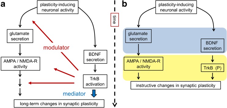
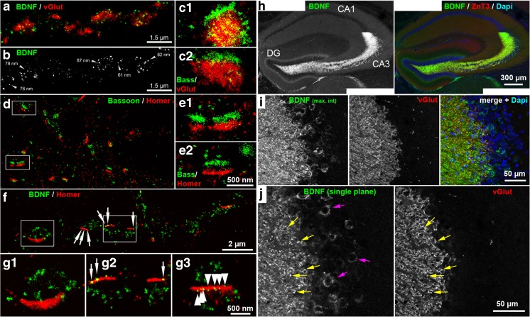
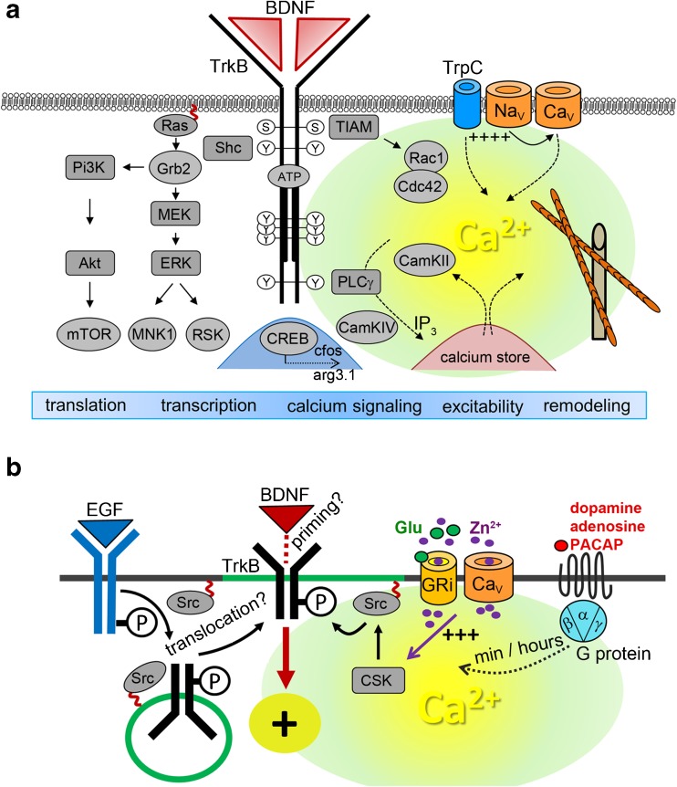
Brain-derived neurotrophic factor (BDNF) is a member of the neurotrophin family of secreted proteins. Signaling cascades induced by BDNF and its receptor, the receptor tyrosine kinase TrkB, link neuronal growth and differentiation with synaptic plasticity. For this reason, interference with BDNF signaling has emerged as a promising strategy for potential treatments in psychiatric and neurological disorders. In many brain circuits, synaptically released BDNF is essential for structural and functional long-term potentiation, two prototypical cellular models of learning and memory formation. Recent studies have revealed an unexpected complexity in the synaptic communication of mature BDNF and its precursor proBDNF, not only between local pre- and postsynaptic neuronal targets but also with participation of glial cells. Here, we consider recent findings on local actions of the BDNF family of ligands at the synapse and discuss converging lines of evidence which emerge from per se conflicting results.

Keywords: Anxiety disorders; BDNF; Long-term potentiation; Signaling; Synaptic localization; Synaptic plasticity; TrkB.


Conflict of interest statement

The authors declare that they have no competing financial interest.

Figures

Fig. 1
Fig. 1
Overview of BDNF signaling. A major source of BDNF in the brain is the excitatory glutamatergic synapse, a principle synapse of synaptic plasticity, learning, and memory. During plasticity-inducing neuronal activity, BDNF and glutamate are released at synapses. a BDNF secretion occurs at a slower timescale than glutamate release. BDNF binds to its receptor TrkB to activate modulatory signaling cascades (see Fig. 3). In presynapses, BDNF-TrkB signaling enhances neurotransmitter release. On postsynaptic sides, BDNF/TrkB signaling increases the function or open probability of ionotropic glutamate receptors. Furthermore, it modulates signaling cascades downstream of neuronal excitation. BDNF as a mediator (according to Park and Poo [126]) can directly influence late effects in synaptic plasticity, for instance, local protein synthesis, spine remodeling, or gene transcription. b BDNF as an instructor of synaptic plasticity. Glutamate and BDNF are released within a critical time window (bright blue) and TrkB activation by BDNF serves as an instructive signal for associative postsynaptic long-term potentiation (bright yellow window)
Fig. 2
Fig. 2
Immunodetection of BDNF in cultured hippocampal neurons and in the hippocampus of the mouse. a, b dSTORM super-resolution images with a resolution of ~20 nm. Immunoreactivity of BDNF and presynaptic vesicular glutamate transporter (vGluT) is shown. Single BDNF-containing granules are located within the vGlut + area, representing the glutamatergic presynapse. b Black-white presentation of the BDNF granules from (a). Single vesicles with a dense BDNF label are pointed out by arrows. These granules have a diameter in the range of 60–90 nm. c 1,2 (details) Presynaptic Bassoon bar structures and vGlut + disks. d Presynaptic Bassoon and postsynaptic Homer1 clusters shown as juxtaposed synaptic bar structures (mouse hippocampal neurons cultured for 35 days). e 1,2 Homer and Bassoon form a bar-like synaptic scaffold structure. f, g BDNF localization at postsynaptic bar structures (DIV 30). BDNF+ vesicles accumulate in juxtaposed position to Homer + postsynaptic bars (white arrows). g 1–3 Some BDNF + granules overlap with the postsynaptic bars. g 3 Multiple (nine) small BDNF + vesicles are aligned within a postsynaptic, Homer + bar (arrows) (a–g : taken from Andreska et al. [8]). h Anti-BDNF immunoreactivity in the hippocampus of the mouse (8-week old, confocal microscopy). Note the overlap with ZnT3 (zinc transporter 3), a protein with high abundance in presynaptic mossy fiber terminals. DAPI labels cell nuclei. i, j BDNF immunoreactivity is pronounced in vGlut + mossy fiber terminals and in somatic areas of CA3 pyramidal neurons. i Maximum intensity projection; j single confocal plane (h–j : performed by M. S. & R.B.)
Fig. 3
Fig. 3
a Overview of BDNF/TrkB signaling in neuronal differentiation and synaptic plasticity. b Activation of TrkB in the absence of neurotrophins. Details are given in the main text. Abbreviations: Akt (protein kinase B), arg3.1 activity-regulated gene 3.1 protein homolog (Arc), BDNF brain-derived neurotrophic factor, CamK Ca2+/calmodulin-dependent protein kinase, Ca V voltage-gated calcium channel, Cdc42 GTPase cell division control protein 42, cfos transcription factor cFos, CREB transcription factor cAMP response element-binding protein, CSK C-terminal Src kinase, ERK extracellular signal regulated kinase, Grb2 growth factor receptor bound protein 2, GRi ionotropic glutamate receptors, IP 3 inositol 1,4,5-trisphosphate, MEK mitogen-activated protein kinase kinase, MNK mitogen-activated protein kinase-interacting kinase, mTOR mechanistic target of rapamycin, Na V voltage-gated sodium channel, PACAP pituitary adenylate cyclase-activating peptide, Pi3K phosphatidylinositol 3-kinase, PLC phospholipase C, Rac GTPase Ras-related C3 botulinum toxin substrate, Ras GTPase rat sarcoma, RSK ribosomal S6 kinase, Shc Src homologous and collagen-like protein, Src Src family of protein tyrosine kinases (SFKs, e.g., Fyn), TIAM T cell lymphoma invasion and metastasis-inducing protein, TrkB tropomyosin-receptor-kinase B, TrpC canonical transient receptor potential channel, Zn 2+ zinc ions
Fig. 4
Fig. 4
Models of synaptic BDNF signaling. a Functional antagonism of BDNF isoforms. In this model, BDNF potentiates the strength of synaptic transmission (plus sign) through TrkB receptors, while under different stimulation conditions, postsynaptic p75NTR receptors are activated by proBDNF to decrease the strength of synaptic transmission (minus sign). Mature BDNF is either secreted or formed by extracellular cleavage from proBDNF, as indicated by the enzymatic scissor. BDNF may be secreted from pre- and/or postsynaptic sources. b Autocrine and paracrine BDNF signaling to increase the strength of synaptic transmission. Presynaptic and postsynaptic TrkB receptors are activated by mature BDNF. TrkB-PLCγ signaling mediates fast BDNF functions and interacts with other phosphorylation cascades to mediate temporally delayed effects, such as structural LTP. BDNF derives from presynaptic and postsynaptic sources and acts autocrine and anterograde. Under certain circumstances, BDNF might act as a potent retrograde messenger. The model includes that TrkB signaling cascades activate synaptic ER calcium stores. The model combines converging lines of evidence at the Schaffer collateral–CA1 synapse. c BDNF signaling at the mossy fiber synapse. This model is focused on anterograde BDNF release. High amounts of BDNF are stored in the presynaptic mossy fiber terminal. Cleaved and uncleaved BDNF isoforms are found in this synapse. Autocrine activation of presynaptic TrkB receptors is involved in presynaptic LTP. BDNF can also excite CA3 pyramidal neurons, either through IP3-mediated ER calcium release and subsequent activation of canonical transient receptor potential (TrpC) channels or fast depolarization (triple plus signs in red). Zinc ions (Zn2+) are co-released with glutamate from mossy fiber synapses, enter CA3 pyramidal neurons through ion channels, and mediate postsynaptic TrkB transactivation. BDNF signaling mechanisms at this synapse might change during development. There is evidence that BDNF plays an important role in feedforward signaling through local interneurons. The temporal and spatial aspects of the indicated signaling mechanisms are unclear. d BDNF signaling at the tripartite synapse in the perirhinal cortex. Secreted proBDNF is taken up by peri-synaptic glial cells through p75NTR and is internalized and recycled as mature BDNF for LTP maintenance. The locus of proBDNF processing is not known

References

    1. Acsady L, Kamondi A, Sik A, Freund T, Buzsaki G. GABAergic cells are the major postsynaptic targets of mossy fibers in the rat hippocampus. The Journal of neuroscience : the official journal of the Society for Neuroscience. 1998;18:3386–3403. - PMC - PubMed
    1. Aicardi G, Argilli E, Cappello S, Santi S, Riccio M, Thoenen H, Canossa M (2004) Induction of long-term potentiation and depression is reflected by corresponding changes in secretion of endogenous brain-derived neurotrophic factor. Proceedings of the National Academy of Sciences of the United States of America 101: 15788–15792 - PMC - PubMed
    1. Aid T, Kazantseva A, Piirsoo M, Palm K, Timmusk T. Mouse and rat BDNF gene structure and expression revisited. J Neurosci Res. 2007;85:525–535. doi: 10.1002/jnr.21139. - DOI - PMC - PubMed
    1. Altar CA, Cai N, Bliven T, Juhasz M, Conner JM, Acheson AL, Lindsay RM, Wiegand SJ. Anterograde transport of brain-derived neurotrophic factor and its role in the brain. Nature. 1997;389:856–860. doi: 10.1038/39885. - DOI - PubMed
    1. Amaral MD, Pozzo-Miller L. BDNF induces calcium elevations associated with IBDNF, a nonselective cationic current mediated by TRPC channels. J Neurophysiol. 2007;98:2476–2482. doi: 10.1152/jn.00797.2007. - DOI - PMC - PubMed

Publication types

Substances