. 2017 Dec;33(6):557-573.
doi: 10.1007/s10565-017-9390-0. Epub 2017 Mar 9.

Proteasome inhibitors against amelanotic melanoma

Affiliations

Proteasome inhibitors against amelanotic melanoma

Justyna Sidor-Kaczmarek et al. Cell Biol Toxicol. 2017 Dec.

Abstract

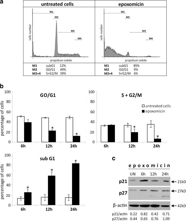
The incidence of malignant melanoma, the most aggressive skin cancer, is increasing constantly. Despite new targeted therapies, the prognosis for patients with metastatic disease remains poor. Thus, there is a need for new combinational treatments, and antineoplastic agents potentially valuable in this approach are inhibitors of the ubiquitin-proteasome system (UPS). In this work, we analyze the cytotoxicity mechanisms of proteasome inhibitors (MG-132, epoxomicin, and lactacystin) in a specific form of melanoma which does not synthesize melanin-the amelanotic melanoma (Ab cells). We found that the most cytotoxic of the compounds tested was epoxomicin. Caspase-9 activation as well as cytochrome C and AIF release from mitochondria indicated that exposure to epoxomicin induced the mitochondrial pathway of apoptosis. Epoxomicin treatment also resulted in accumulation of Bcl-2 family members-proapoptotic Noxa and antiapoptotic Mcl-1, which were postulated as the targets for bortezomib in melanoma. Inhibition of caspases by BAF revealed that cell death was partially caspase-independent. We observed no cell cycle arrest preceding the apoptosis of Ab cells, even though cdk inhibitors p21Cip1/Waf1 and p27Kip1 were up-regulated. The cell cycle was blocked only after inactivation of caspases by the pan-caspase inhibitor BAF. In summary, this is the first study exploring molecular mechanisms of cell death induced by epoxomicin in melanoma. We found that Ab cells died on the mitochondrial pathway of apoptosis and also partially by the caspase-independent way of death. Apoptosis induction was fast and efficient and was not preceded by cell cycle arrest.

Keywords: Apoptosis; Cancer; Melanoma; Proteasome inhibitors.


Figures

Fig. 1
Fig. 1
Cytotoxic effect of proteasome inhibitors on Ab melanoma cells. Cell viability was determined by XTT assay and expressed as a percentage of the viability of untreated cells. Cells were exposed to increasing concentrations of epoxomicin, MG-132, or lactacystin for 12 (a) and 24 (b) hours or to 0.5 μM epoxomicin, 5 μM MG-132, or 10 μM lactacystin for up to 72 h (c). An asterisk (*) indicates a statistically significant difference between various proteasome inhibitor concentrations; p < 0.05, Kruskal-Wallis test. d Inhibition of chymotrypsin-like (ChT-L) proteasome activity in Ab melanoma cells. Cells were exposed to 0.5 μM epoxomicin, 5 μM MG-132, or 10 μM lactacystin for 2 h, and proteasome activity was measured using a luminescent assay. The data represent means ± SD of three independent experiments
Fig. 2
Fig. 2
Proteasome inhibition-induced apoptotic cell death changes in Ab melanoma cells. Cells were exposed to 0.5 μM epoxomicin for 6, 12, and 24 h. a Hoechst 33342 staining of Ab cells after 24 h of treatment. Fluorescence of apoptotic nuclei is more intense than in viable cells. Chromatin condensation and nucleus fragmentation are prominent after treatment (arrow). b Apoptotic plasma membrane changes in Ab cells. Cells were stained with FITC-conjugated annexin V (An) and propidium iodide (PI) to estimate early (An+/PI−) and late apoptotic (An+/PI+) cells. Representative dot plots from flow cytometry analysis results are shown. c Increase in early (An+/PI−) and late (An+/PI+) apoptotic Ab melanoma cells after exposure to epoxomicin. The data represent means ± SD of three independent experiments. An asterisk (*) indicates a statistically significant increase in apoptotic cells in comparison to untreated cells, p < 0.05, Kruskal-Wallis test
Fig. 3
Fig. 3
Epoxomicin-induced mitochondrial pathway of apoptosis and HSP expression in Ab melanoma cells. Cells were exposed to 0.5 μM epoxomicin for 6, 12, and 24 h. a Immunoblot analysis of caspase 3 and 9 activation and expression of Mcl-1, Noxa, and HSP proteins in Ab cells in response to epoxomicin. β-Actin was used as a control of the equal protein loading. UN untreated cells. The densitometric ratio of band intensity is shown. b Double-labeled immunofluorescent staining revealing cytochrome C and AIF release from mitochondria in Ab cells treated with epoxomicin for 6 h. Upper panel cytochrome C (green) and mitochondria (red), an arrow indicates the membrane blebbing specific for apoptotic cell death; lower panel: AIF (green) and cell nucleus (red)
Fig. 4
Fig. 4
Epoxomicin-induced apoptosis of Ab melanoma cells only partially relies on caspase activation. Ab cells were treated with 0.5 μM epoxomicin alone or in the presence of 50 μM pan-caspase inhibitor BAF for 24 h. a Cell viability was measured by an XTT assay and calculated as a percentage of the viability of untreated cells. b Cells were stained with FITC-conjugated annexin V (An) and propidium iodide (PI) to estimate live cells (An−/PI−), early (An+/PI−) and late apoptotic (An+/PI+) cells. In a and b, the data represent the means ± SD of three independent experiments; asterisk (*) indicates statistically significant difference between BAF 0 μM and BAF 50 μM, Mann-Whitney U test, p < 0.01 (a) or p < 0.05 (b). c Ab cells were treated with epoxomicin alone or in the presence of BAF and stained with Hoechst 33342. Arrows indicate the nucleus fragmentation in the absence of BAF (upper panel, BAF 0 μM) and chromatin condensation, but no nucleus fragmentation in the presence of BAF (lower panel, BAF 50 μM). d Cell cycle distribution was determined in PI-stained Ab cells by flow cytometry. Representative histograms are shown. The percentage of cells with fragmented DNA (subG1, M1) and in the G2/M (M4) phase of the cell cycle are indicated below the corresponding histograms
Fig. 5
Fig. 5
Ab melanoma cell cycle analysis and changes in the cdk (cell cycle-dependent kinases) inhibitors p21Cip1/Waf1 and p27Kip1 after epoxomicin treatment. Cells were exposed to 0.5 μM epoxomicin for up to 24 h. a Representative histograms of cell cycle analysis of cells exposed to epoxomicin for 24 h. DNA content was estimated by propidium iodide staining, and cell cycle distribution was assessed by flow cytometry. b Time-dependent decrease in G0/G1 and S + G2/M phases and accumulation of destroyed cells in subG1. Data represent means ± SD of three independent experiments. Asterisks (*) indicate statistically significant difference between untreated and epoxomicin-treated cells (p < 0.05), Kruskal-Wallis test. c Immunoblot analysis of p21Cip1/Waf1 and p27Kip1 up-regulation in Ab cells treated with 0.5 μM epoxomicin. β-actin was used as a control of the equal protein loading. UN-untreated cells. The densitometric ratio of band intensity is shown
Fig. 6
Fig. 6
Principal mechanisms of action of proteasome inhibitors in cancer cells including melanoma. Principal molecular mechanisms underlying cytotoxic effect of proteasome inhibition involve the following: (i) up-regulation of pro-apoptotic Bcl-2 family proteins, (ii) stabilization of CDK inhibitors, (iii) p53 stabilization, (iv) endoplasmic reticulum (ER) stress, (v) decreased NF-κB signaling, and (vi) oxidative stress (accumulation of ROS). These changes result in cell cycle arrest and apoptosis in cancer cells. Inhibition of proteasome activity increases expression of pro-apoptotic BH3-only members of Bcl-2 family, including Noxa, and in some models down-regulates anti-apoptotic factors such as Bcl-2 and Mcl-1. Enhanced pro-apoptotic signaling promotes the assembly of Bax-Bak oligomers in mitochondrial outer membrane, cytochrome C and AIF release to cytosol, apoptosome formation and finally activation of caspases. Apoptotic signals are augmented by p53, which is activated by ROS-induced DNA damage and further stabilized by blocking its proteasomal degradation after ubiquitination by Mdm2. Accumulated p53 activates expression of its target genes: Puma, Noxa, and Bax. p53 also arrests cell cycle by inducing expression of CDK inhibitor p21. CDK inhibitors p21 and p27 are also directly up-regulated by proteasome inhibitors, which prevent their proteasomal degradation after ubiquitination by Skp-2. Another mechanism promoting apoptosis is down-regulation of NF-κB signaling. Inhibition of proteasome prevents degradation of inhibitory protein-IκB and subsequent activation and translocation of NF-κB to the nucleus. Aggregation of misfolded proteins resulting from proteasome inhibition triggers ER stress and defense mechanism called unfolded protein response (UPR). Aggregated proteins activate ER transmembrane proteins PERK, IRE1α, and transcription factor ATF6 by their homodimerization and proteolytical processing. ER stress also triggers HSF-1 trimerization and binding to HSP promoters. This mechanism initially protects the cell by increased expression of protein chaperones HSP70, HSP27, and BiP/Grp78. Overexpressed HSP70 and HSP27 are inhibitors of apoptosis. HSP70 inhibits assembly of Bax oligomers, prevents recruitment of procaspase-9 to apoptosome, and blocks nuclear translocation of AIF and chromatin condensation. HSP27 associates with cyt C and inhibits the formation of apoptosome, decreases ROS content, and targets IκB for ubiquitination. When overwhelmed, UPR triggers apoptosis by activation of CHOP. Elements examined in the present study are shown in red. → activation, ⊣ inhibition. AIF apoptosis-inducing factor, Apaf-1 apoptotic protease activating factor 1, ATF activating transcription factor, Bak Bcl-2 antagonist/killer-1, Bax Bcl-2-associated X protein, Bcl-2 B cell lymphoma 2, Bik BCL2 interacting killer, Bim Bcl-2-like protein 11, BiP/Grp78 binding immunoglobulin protein/78 kDa glucose-regulated protein, CDK cyclin-dependent kinase, CHOP CCAAT/enhancer-binding protein homologous protein, eIF2a eukaryotic initiation factor 2, IκB inhibitory protein κB, HSF-1 heat-shock transcription factor 1, IRE1α inositol-requiring enzyme 1, Mcl-1 myeloid cell leukemia 1, NF-κB nuclear factor κB, PERK ER-resident protein kinase, Puma p53-up-regulated modulator of apoptosis, ROS reactive oxygen species, Skp-2 S-phase kinase-associated protein 2, XBP1 X-box binding protein 1

Similar articles

Cited by

References

    1. Adams J, Kauffman M. Development of the proteasome inhibitor Velcade (bortezomib) Cancer Investig. 2004;22:304–311. doi: 10.1081/CNV-120030218. - DOI - PubMed
    1. Amann VC, Ramelyte E, Thurneysen S, Pitocco R, Bentele-Jaberg N, Goldinger SM, et al. Developments in targeted therapy in melanoma. Eur J Surg Oncol. 2016:1–13. - PubMed
    1. Amschler K, Schön MP, Pletz N, Wallbrecht K, Erpenbeck L, Schön M. NF-kappa B inhibition through proteasome inhibition or IKKbeta blockade increases the susceptibility of melanoma cells to cytostatic treatment through distinct pathways. J Invest Dermatol. 2010;130:1073–1086. doi: 10.1038/jid.2009.365. - DOI - PubMed
    1. Berenson A, Vardanyan S, David M, Wang J, Harutyunyan NM, Gottlieb J, et al. Outcomes of multiple myeloma patients receiving bortezomib, lenalidomide, and carfilzomib. Ann Hematol. 2016 (ahead of print) - PubMed
    1. Bomirski A, Słominski A, Bigda J. The natural history of a family of transplantable melanomas in hamsters. Cancer Metastasis Rev. 1988;7:95–118. doi: 10.1007/BF00046481. - DOI - PubMed

Publication types

Substances