. 2018 Mar;28(2):264-273.
doi: 10.1111/bpa.12505. Epub 2017 Mar 24.

Overlapping but distinct TDP-43 and tau pathologic patterns in aged hippocampi

Affiliations

Overlapping but distinct TDP-43 and tau pathologic patterns in aged hippocampi

Vanessa D Smith et al. Brain Pathol. 2018 Mar.

Abstract

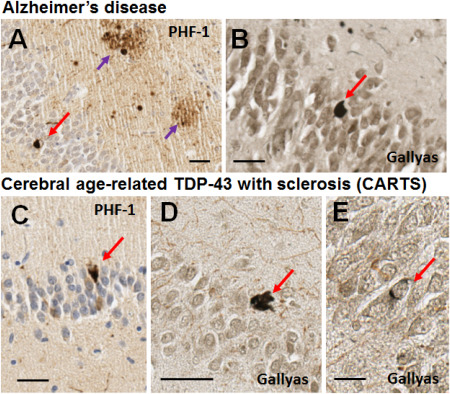
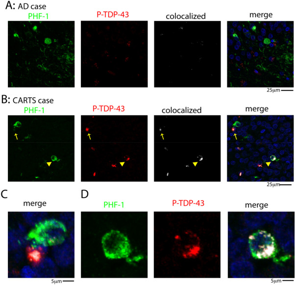
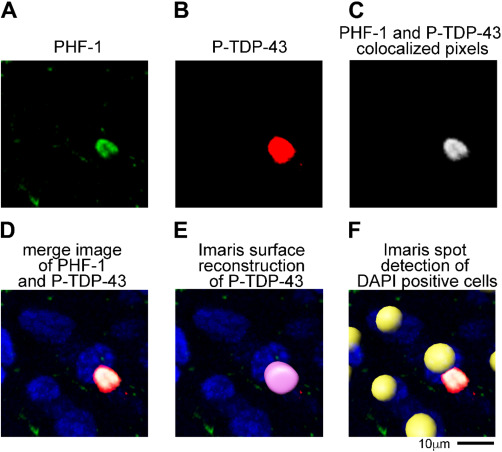
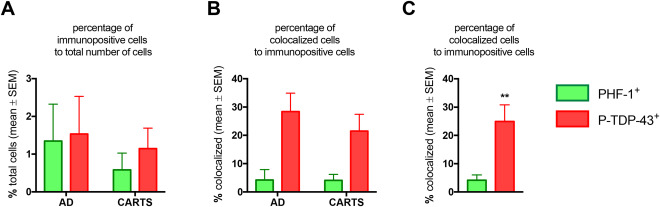
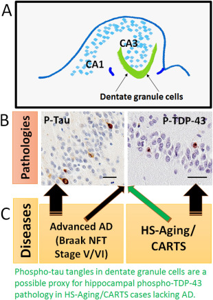
Intracellular proteinaceous aggregates (inclusion bodies) are almost always detectable at autopsy in brains of elderly individuals. Inclusion bodies composed of TDP-43 and tau proteins often coexist in the same brain, and each of these pathologic biomarkers is associated independently with cognitive impairment. However, uncertainties remain about how the presence and neuroanatomical distribution of inclusion bodies correlate with underlying diseases including Alzheimer's disease (AD). To address this knowledge gap, we analyzed data from the University of Kentucky AD Center autopsy series (n = 247); none of the brains had frontotemporal lobar degeneration. A specific question for this study was whether neurofibrillary tangle (NFT) pathology outside of the Braak NFT staging scheme is characteristic of brains with TDP-43 pathology but lacking AD, that is those with cerebral age-related TDP-43 with sclerosis (CARTS). We also tested whether TDP-43 pathology is associated with comorbid AD pathology, and whether argyrophilic grains are relatively likely to be present in cases with, vs. without, TDP-43 pathology. Consistent with prior studies, hippocampal TDP-43 pathology was associated with advanced AD - Braak NFT stages V/VI. However, argyrophilic grain pathology was not more common in cases with TDP-43 pathology in this data set. In brains with CARTS (TDP-43[+]/AD[-] cases), there were more NFTs in dentate granule neurons than were seen in TDP-43[-]/AD[-] cases. These dentate granule cell NFTs could provide a proxy indicator of CARTS pathology in cases lacking substantial AD pathology. Immunofluorescent experiments in a subsample of cases found that, in both advanced AD and CARTS, approximately 1% of dentate granule neurons were PHF-1 immunopositive, whereas ∼25% of TDP-43 positive cells showed colocalized PHF-1 immunoreactivity. We conclude that NFTs in hippocampal dentate granule neurons are often present in CARTS, and TDP-43 pathology may be secondary to or occurring in parallel with tauopathy.

Keywords: FTLD; HS-aging; colocalization; hippocampal sclerosis; hippocampus; oldest-old.


Figures

Figure 1
Figure 1
Brightfield photomicrographs depict representative PHF‐1 immunohistochemistry for phospho‐tau (A,C) and Gallyas silver stain (B,D,E) in dentate granule neurons in Alzheimer's disease and CARTS. The Alzheimer's disease case (A,B) was a male with APOE ε 3/4 alleles, who died at age 85. Neuropathology showed Braak NFT Stage VI with CERAD neuritic plaque score of “moderate”. No TDP‐43 pathology was observed. The CARTS case (C,D,E) was a female with APOE ε 2/3 alleles, who died at age 90. Thal Aβ stage was 1 (very few neocortical plaques detected with Aβ immunohistochemistry), CERAD neuritic plaque score of “none”, and Braak NFT stage I. There was substantial TDP‐43 pathology in the hippocampus and subiculum. In both cases, NFTs were detected in dentate granule neurons with PHF‐1 antibody, and with the Gallyas silver impregnation technique. Some of the NFTs stained with silver impregnation were dark fibrillar NFTs (D) whereas some were more subtle (E). Red arrows show NFTs, purple arrows show tau‐positive neuritic plaques. Scale bar = 30 μM (A,B), 40 μM (C), 50 μM (D), and 15 μM (E).
Figure 2
Figure 2
Double‐label immunofluorescent images for tau (PHF‐1) tangles and P‐TDP‐43 pathology in hippocampal dentate granule neurons. Representative examples are shown of PHF‐1 and P‐TDP‐43 staining in an AD case (A) and a CARTS case (B). Arrow in (B) shows a P‐TDP‐43 inclusion in higher magnification in the merged photomicrograph indicating the PHF‐1 (green) and P‐TDP‐43 (red) signals are not colocalized (C). By contrast, the arrowhead in (B) shows a PHF‐1 positive cell that is colocalized with P‐TDP‐43, as shown at a higher magnification in (D).
Figure 3
Figure 3
Cell counting and colocalization method used Imaris 3D microscopic data analysis software on confocal Z‐stack images. (A) PHF‐1 was thresholded to exclude pixels detected in the no PHF‐1 primary antibody control samples. (B) P‐TDP‐43 was similarly thresholded to exclude pixels detected in the no P‐TDP‐43 primary antibody control samples. (C) Using the Imaris Coloc function a new channel was generated that corresponded to the pixels (after thresholding) that were in present in both the PHF‐1 and P‐TDP‐43 channel. The merged image of the channels is shown in (D). (E) A surface reconstruction was generated for the PHF‐1 and P‐TDP‐43 to unbiasedly count the number of cell (PHF‐1) and inclusions (P‐TDP‐43) in each image. Note that the PHF‐1 staining in (A) was below the size threshold to be counted as a cell, thus no surface was rendered for the PHF‐1 inclusion. (F) Imaris spot detection function was used to count the total number of DAPI positive cells in images. The yellow spheres in the picture highlight the detection of individual DAPI positive cells using the spot detection function.
Figure 4
Figure 4
Results of counts and colocalization of PHF‐1 and P‐TDP‐43 immunoreactive profiles in dentate granule neurons, in advanced AD (Braak NFT stages) and CARTS cases. This is the result of analyses of a subsample of 6 cases with 2 slides/case analyzed, comprising 14 200 dentate granule neurons from CARTS cases and 11 952 cells from AD cases. Panel A shows the overall percentage of immunopositive cells. Note that approximately 1% of cells are positive for PHF‐1 and P‐TDP‐43, with trend for more PHF‐1 immunoreactive cells in the dentate granule cell layer from AD cases. Panel B shows the percentage of cells that are colocalized among those that are immunopositive. Panel C shows that in both AD and CARTS, a higher percentage (∼25%) of cells that were TDP‐43 immunopositive were also PHF‐1 immunopositive, in comparison with the colocalization among the PHF‐1 immunopositive cells in terms of P‐TDP‐43 immunopositivity (4%) **P = 0.007.
Figure 5
Figure 5
Hippocampal dentate granule NFTs are associated with advanced AD and also with the presence of TDP‐43 pathology in low Braak stage (CARTS) cases. Schematic depiction of hippocampal formation (A) to show how the pathologies studied in the dentate granule neurons (B) are correlated with different common brain diseases (C). Both phospho‐Tau and phospho‐TDP‐43 pathologies commonly occur in both advanced AD and in CARTS. This implies that disparate upstream causal factors may lead to common downstream pathologic pathways. The appearance of dentate granule NFTs in cases without AD pathology may serve as a useful proxy for indicating CARTS (green arrow). Both of these particular brightfield photomicrographs were taken from the same CARTS case, counterstained with hematoxylin; scale bars = 40 μM.

References

    1. Abisambra JF, Scheff S (2014) Brain injury in the context of tauopathies. J Alzheimer's Dis 40:495–518. - PubMed
    1. Amador‐Ortiz C, Ahmed Z, Zehr C, Dickson DW (2007) Hippocampal sclerosis dementia differs from hippocampal sclerosis in frontal lobe degeneration. Acta Neuropathol 113:245–252. - PMC - PubMed
    1. Amador‐Ortiz C, Lin WL, Ahmed Z, Personett D, Davies P, Duara R et al (2007) TDP‐43 immunoreactivity in hippocampal sclerosis and Alzheimer's disease. Ann Neurol 61:435–445. - PMC - PubMed
    1. Aoki N, Murray ME, Ogaki K, Fujioka S, Rutherford NJ, Rademakers R et al (2015) Hippocampal sclerosis in Lewy body disease is a TDP‐43 proteinopathy similar to FTLD‐TDP Type A. Acta Neuropathol 129:53–64. - PMC - PubMed
    1. Arnold SJ, Dugger BN, Beach TG (2013) TDP‐43 deposition in prospectively followed, cognitively normal elderly individuals: correlation with argyrophilic grains but not other concomitant pathologies. Acta Neuropathol 126:51–57. - PMC - PubMed

Publication types

MeSH terms