. 2017 Mar 10:8:14706.
doi: 10.1038/ncomms14706.

Adipocytes promote malignant growth of breast tumours with monocarboxylate transporter 2 expression via β-hydroxybutyrate

Affiliations

Adipocytes promote malignant growth of breast tumours with monocarboxylate transporter 2 expression via β-hydroxybutyrate

Chun-Kai Huang et al. Nat Commun. .

Abstract

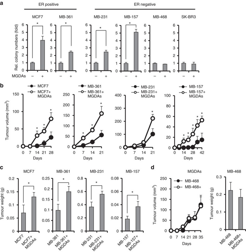
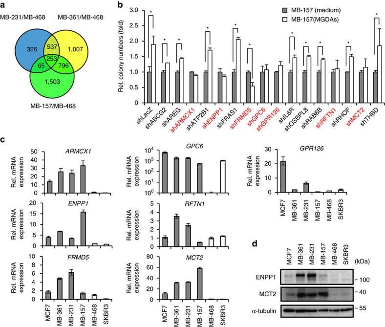
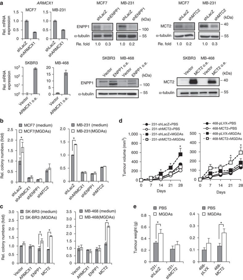
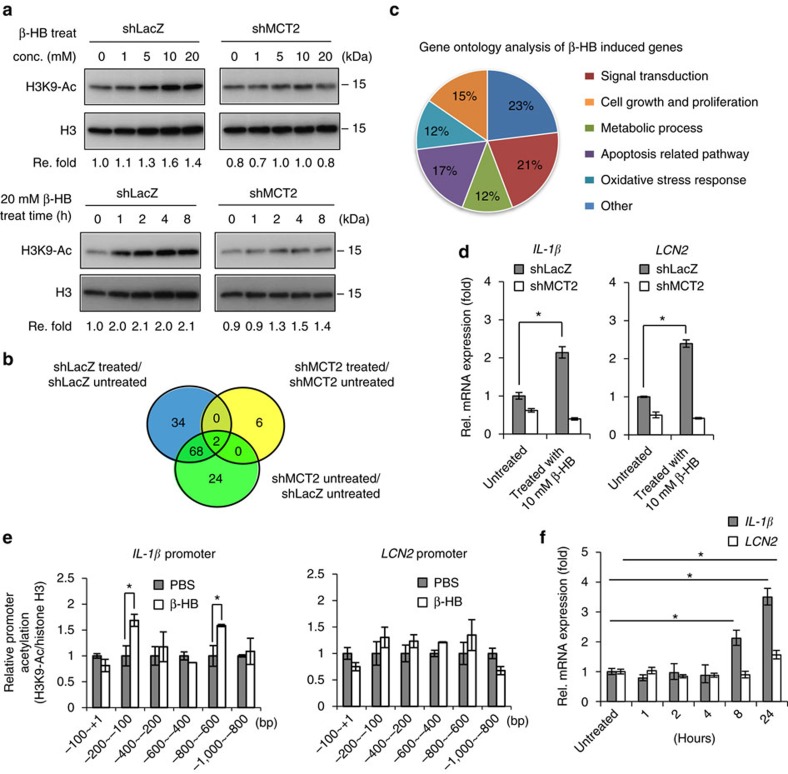
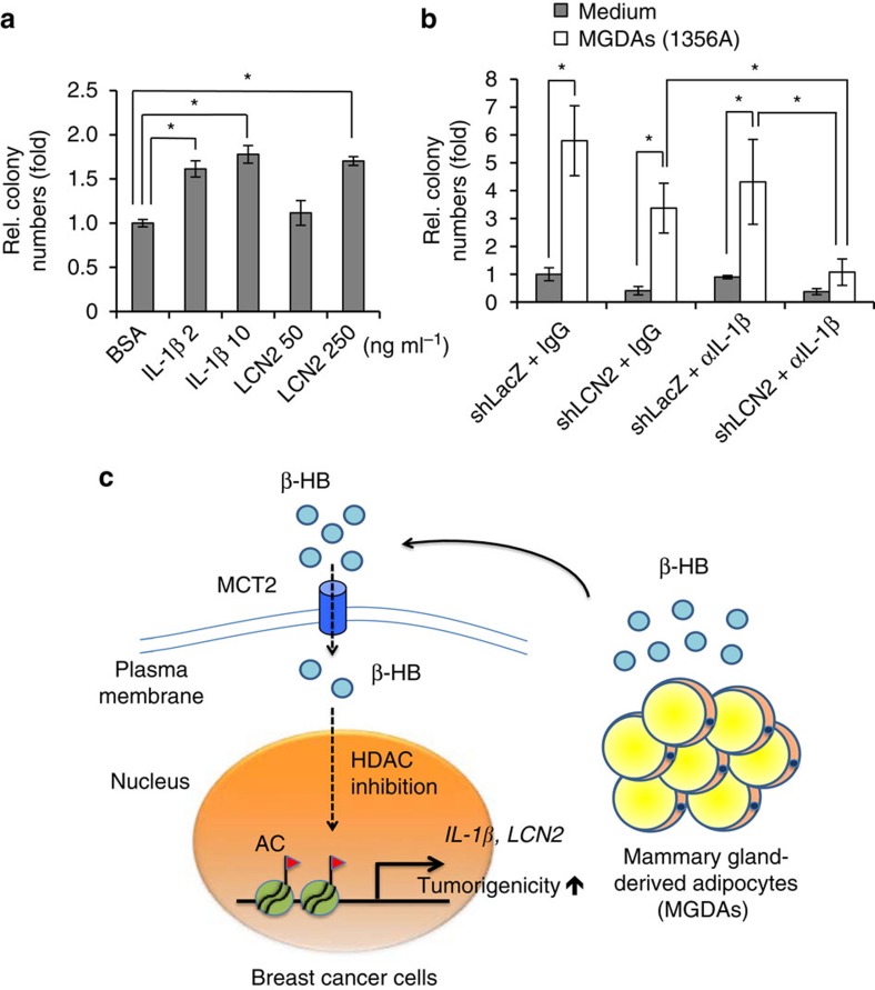
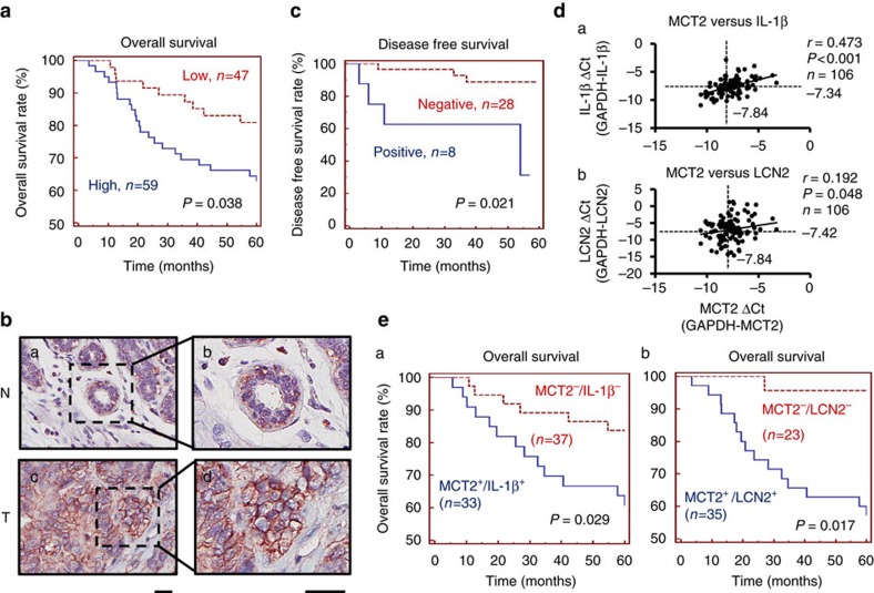
Adipocytes are the most abundant stromal partners in breast tissue. However, the crosstalk between breast cancer cells and adipocytes has been given less attention compared to cancer-associated fibroblasts. Here we find, through systematic screening, that primary mammary gland-derived adipocytes (MGDAs) promote growth of breast cancer cells that express monocarboxylate transporter 2 (MCT2) both in vitro and in vivo. We show that β-hydroxybutyrate is secreted by MGDAs and is required to enhance breast cancer cells malignancy in vitro. Consistently, β-hydroxybutyrate is sufficient to promote tumorigenesis of a mouse xenograft model of MCT2-expressing breast cancer cells. Mechanistically we observe that upon co-culturing with MGDAs or treatment with β-hydroxybutyrate, breast cancer cells expressing MCT2 increase the global histone H3K9 acetylation and upregulate several tumour-promoting genes. These results suggest that adipocytes promote malignancy of MCT2-expressing breast cancer via β-hydroxybutyrate potentially by inducing the epigenetic upregulation of tumour-promoting genes.


Conflict of interest statement

The authors declare no competing financial interests.

Figures

Figure 1
Figure 1. Mammary gland-derived adipocytes promote tumour growth in several breast cancer cell lines.
(a) Soft agar colony formation assays using a panel of breast cancer cell lines co-cultured with MGDAs. (b) Tumour growth assays in NOD/SCID/γnull mice. Breast cancer cells were subcutaneously injected in both flanks of each mouse w/ or w/o MGDAs and tumour volumes were measured every 7 days. Four mice (n=4) were used for each group. (c) Co-injection of MGDAs in xenograft models increased the tumour weights in several breast cancer cell lines. (d) MGDAs co-injection failed to enhance the tumour growth of MDA-MB-468 cells in mouse tumorigenicity assays. The experiment in a was performed in technical triplicate and repeated at least twice with similar results. In ad, data show means±s.d. *P<0.05 (Student's t-test).
Figure 2
Figure 2. Identification of membrane proteins involved in MGDAs-mediated enhancement of tumorigenic activity.
(a) Summary of cDNA microarray analyses. In all, 253 genes were identified with at least twofold upregulation in MDA-MB-231, MDA-MB-361 and MDA-MB-157 compared to MDA-MB-468 cells. (b) RNAi knockdown screening of candidate genes involved in MGDAs-mediated tumorigenic activity promotion in MDA-MB-157 cells by soft agar colony formation assay. shLacZ was used as a knockdown control. (c) Expression profiles of ARMCX1, ENPP1, FRMD5, GPC6, GPR126, RFTN1 and MCT2 in six different breast cancer cell lines. The experiment was performed in technical triplicate. (d) Western blot analyses reconfirmed the expression profiles of ENPP1 and MCT2 in breast cancer cell lines (no commercial workable ARMCX1 antibody was found). The experiment in b was performed in technical triplicate and repeated at least twice with similar results. Data show means±s.d. *P<0.05 (Student's t-test).
Figure 3
Figure 3. MCT2 is required for the enhancement of tumorigenic activity associated with MGDAs.
(a) Q-PCR and western blot analyses of ARMCX1, ENPP1 and MCT2 depletion and ectopic overexpression in breast cancer cells. (b) Soft agar colony formation assays showed that depletion of ARMCX1, ENPP1 and MCT2 abrogated the increase of colonies induced by MGDAs co-culture in MCF7 and MDA-MB-231 cells. (c) Soft agar colony formation assays of ARMCX1, ENPP1 and MCT2 overexpressing MDA-MB-468 and SK-BR3 cells w/ or w/o MGDAs co-culture. (d) Tumour growth assays in NOD/SCID/γnull mice. MCT2-depleted MDA-MB-231 and overexpressing MDA-MB-468 cells were subcutaneously injected in both flanks of each mouse w/ or w/o MGDAs. Tumour volumes were measured every 7 days. Four mice (n=4) were used for each group. (e). Tumour weights were increased in the presence of MCT2 after co-injection with MGDAs in mouse tumorigenicity assays. The experiments in b,c were performed in technical triplicate and repeated at least twice with similar results. In be, data show means±s.d. *P<0.05 (Student's t-test).
Figure 4
Figure 4. β-hydroxybutyrate from MGDAs promotes breast cancer progression.
(a) Soft agar colony formation assays of MDA-MB-231 and MDA-MB-468 cells treated with conditioned medium collected from MGDAs culture. The MGDAs-conditioned medium was fractionated by ultrafiltration into <10 kD, 10–50 kD and >50 kD fractions. (b) Depletion or overexpression of MCT2 in MDA-MB-231 or MDA-MB-468 cells either abrogated or enhanced the susceptibility to treatment with fractionated MGDAs-conditioned medium. (c) Secretion levels of β-hydroxybutyrate, lactate and pyruvate in conditioned medium from MGDAs and stromal vascular fraction (SVF) cells were determined by ELISA analyses, respectively. The experiment was performed in technical triplicate. (d) MCT2-depleted MDA-MB-231 and MCT2-overexpressing MDA-MB-468 cells were treated with various doses of β-hydroxybutyrate in soft agar colony formation assays. (e) Tumour growth assays in NOD/SCID/γnull mice. MCT2-depleted MDA-MB-231 and MDA-MB-157 cells were subcutaneously injected in both flank of each mouse, and the mice were administered PBS alone or PBS containing β-hydroxybutyrate (500 mg kg−1) through daily intraperitoneal injection. The tumour volumes were measured every 7 days. Six mice (n=6) were used for each group. The experiments in a,b,d were performed in technical triplicate and repeated at least twice with similar results. In ae, data show means±s.d. *P<0.05 (Student's t-test).
Figure 5
Figure 5. Treatment of β-hydroxybutyrate induces changes of gene expression profiles through epigenetic effects.
(a) The level of H3K9 acetylation in MCT2-depleted MDA-MB-231 cells was assessed upon β-hydroxybutyrate treatment using a variety of doses and times. The experiment was repeated at least twice with similar results. (b) Summary of cDNA microarray analyses. Thirty-four genes were identified with at least 1.5-fold upregulation in β-hydroxybutyrate-treated MDA-MB-231 cells in an MCT2-dependent manner. (c) GO analysis of β-hydroxybutyrate-induced genes. The GO terms were classified into six groups and the percentages were shown. (d) Q-PCR analyses confirmed the expression levels of IL-1β and LCN2 in MDA-MB-231 cells treated with β-hydroxybutyrate. (e) Chromatin immunoprecipitation of acetylated histone H3K9 on IL-1β and LCN2 proximal promoter regions in MDA-MB-231 cells upon 20 mM β-hydroxybutyrate treatment for 1 h. (f) IL-1β and LCN2 expression levels in MDA-MB-231 cells treated with 20 mM β-hydroxybutyrate for various times. The experiments in df were performed in technical triplicate and repeated at least twice with similar results. Data show means±s.d. *P<0.05 (Student's t-test).
Figure 6
Figure 6. IL-1β and LCN2 play important roles in MGDAs-mediated tumorigenicity enhancement.
(a) MDA-MB-231 cells were supplemented with recombinant IL-1β and LCN2 in soft agar colony formation assays. (b). Soft agar colony formation assays showed that knockdown of LCN2 combining IL-1β neutralizing antibody treatment (αIL-1β, 1 μg ml−1) significantly abrogated the increase of colonies induced by MGDAs co-culture with MDA-MB-231 cells. Mouse IgG (1 μg ml−1) was used as a control. The experiments in a,b were performed in technical triplicate and repeated at least twice with similar results. Data show means±s.d. *P<0.05 (Student's t-test). (c) Schematic shows the heterotypic interaction between MGDAs and MCT2-expressing breast cancer cells in the breast tissue microenvironment. MGDAs promote the tumorigenicity of MCT2-expressing breast cancer cells in a paracrine manner. β-hydroxybutyrate secreted from MGDAs can be transported into breast cancer cells via MCT2. Intracellular β-hydroxybutyrate functions as a class I HDAC inhibitor and induces the expression of tumour-promoting genes through epigenetic modification, leading to tumorigenicity enhancement.
Figure 7
Figure 7. Elevated expressions of IL-1β and LCN2 correlate with poor prognosis in breast cancer patients.
(a) Kaplan-Meier analysis showed correlation between cumulative survival and MCT2 expression levels in breast cancer patients. (b) IHC staining of MCT2. The pictures showed the negative (a,b) and positive (c,d) membrane staining of MCT2 in normal and breast cancer tissues, respectively. (Scale bars, 25 μm). (c) Kaplan-Meier survival analysis of patients with MCT2-positive and -negative IHC staining. (d) Correlation analyses of gene expression levels between MCT2, IL-1β and LCN2. MCT2 versus: (a) IL-1β and (b) LCN2. (e) Survival analyses showed (a) MCT2 and IL-1β (b) MCT2 and LCN2 double-positive group versus double-negative group, respectively.

References

    1. Liotta L. A. & Kohn E. C. The microenvironment of the tumour-host interface. Nature 411, 375–379 (2001). - PubMed
    1. Rong S. et al.. Tumorigenicity of the met proto-oncogene and the gene for hepatocyte growth factor. Mol. Cell. Biol. 12, 5152–5158 (1992). - PMC - PubMed
    1. Orimo A. et al.. Stromal fibroblasts present in invasive human breast carcinomas promote tumor growth and angiogenesis through elevated SDF-1/CXCL12 secretion. Cell 121, 335–348 (2005). - PubMed
    1. Tyan S. W. et al.. Breast cancer cells induce cancer-associated fibroblasts to secrete hepatocyte growth factor to enhance breast tumorigenesis. PLoS ONE 6, e15313 (2011). - PMC - PubMed
    1. Chen J. et al.. CCL18 from tumor-associated macrophages promotes breast cancer metastasis via PITPNM3. Cancer cell 19, 541–555 (2011). - PMC - PubMed

Publication types

MeSH terms