. 2017 Mar 10:8:14671.
doi: 10.1038/ncomms14671.

Structural basis of homo- and heterotrimerization of collagen I

Affiliations

Structural basis of homo- and heterotrimerization of collagen I

Urvashi Sharma et al. Nat Commun. .

Abstract

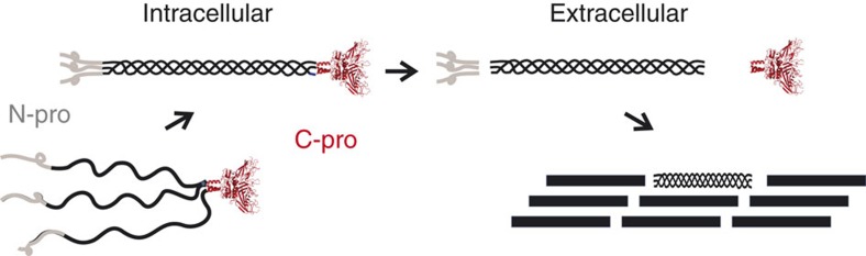
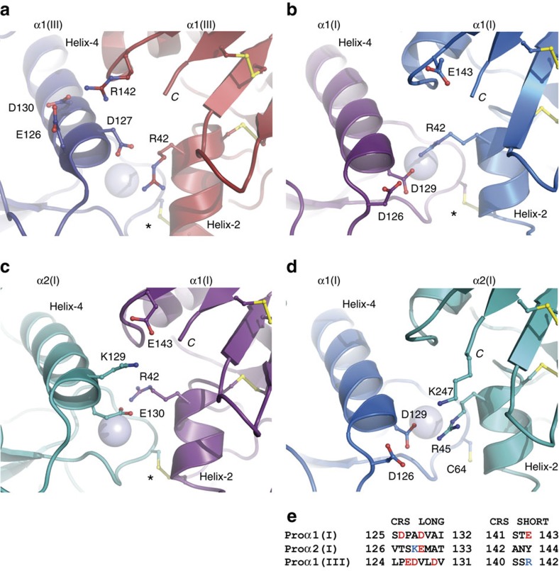
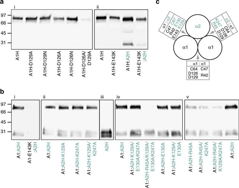
Fibrillar collagen molecules are synthesized as precursors, procollagens, with large propeptide extensions. While a homotrimeric form (three α1 chains) has been reported in embryonic tissues as well as in diseases (cancer, fibrosis, genetic disorders), collagen type I usually occurs as a heterotrimer (two α1 chains and one α2 chain). Inside the cell, the role of the C-terminal propeptides is to gather together the correct combination of three α chains during molecular assembly, but how this occurs for different forms of the same collagen type is so far unknown. Here, by structural and mutagenic analysis, we identify key amino acid residues in the α1 and α2 C-propeptides that determine homo- and heterotrimerization. A naturally occurring mutation in one of these alters the homo/heterotrimer balance. These results show how the C-propeptide of the α2 chain has specifically evolved to permit the appearance of heterotrimeric collagen I, the major extracellular building block among the metazoa.


Conflict of interest statement

The authors declare no competing financial interests.

Figures

Figure 1
Figure 1. Roles of the procollagen C-propeptides.
Trimerization of the procollagen C-propeptides initiates intracellular assembly of the procollagen molecule while extracellular proteolytic cleavage of the N- and C-propeptides controls collagen fibril formation.
Figure 2
Figure 2. 3D structure of homo-CPI.
(a) View from the side showing the stalk, base and petal regions. Each chain is represented in a different colour, with bound Ca2+ ions as light blue spheres and the Cl ion in green. Disulfide bonds are in yellow. (b) View from the top showing a structural alignment of the homo-CPI structure (in colour) on the 3.3 Å structure of CPIII (PDB code 4AK3; in grey). While overall the two structures are well aligned, there is a shift in orientation of helix-4 (highlighted for one chain from each structure).
Figure 3
Figure 3. Interaction interfaces in different C-propeptide trimers.
(a) Charged residues at the A:B chain interface in the 1.7 Å structure of CPIII (PDB code 4AE2). Each chain is shown in a different colour, as in Bourhis et al.. (b) Charged residues at the inter-chain interface in homo-CPI. Chains are coloured as in Fig. 2. Note the large conformational change in the side-chain of residue Arg42 between CPIII and homo-CPI. (c,d) Models of the two inter-chain interfaces in hetero-CPI involving the α2(I) chain. Chains are coloured as in Fig. 2, with the chain in deep teal (greenish blue) replaced by the α2(I) chain. C denotes the C-terminus of each chain and * indicates the position of the inter-chain disulfide bond, which is absent in d leaving the free cysteine Cys64. (e) Sequence alignment in the CRS region for CPI (α1 and α2 chains) and CPIII. See also Supplementary Movies 1,2,3,4.
Figure 4
Figure 4. Effects of site-directed mutations on the trimerization of homo-CPI and hetero-CPI.
Proteins secreted into the culture medium after site-directed mutagenesis of the residues indicated were detected by western blotting using the N-terminal His-tag present on either or both the α1(I) and α2(I) chains, as indicated by H (for example, A1H means His-tagged α1 chain). The α2(I) chain is indicated by the green/blue colour. Gels were run in non-reducing conditions, where trimers migrate at 85 kDa and monomers at 30 kDa. (a) Controls and mutants using the His-tagged α1(I) chain. (b) Controls and mutants using the non-tagged α1(I) chain and the His-tagged α2(I) chain. (c) Schematic view down the axis of a C-propeptide heterotrimer showing interacting residues at the homo- and hetero-chain interfaces. Same colour coding throughout. Data shown are representative of triplicate (a) or duplicate (b) biological replicates.

References

    1. Hynes R. O. & Naba A. Overview of the matrisome-an inventory of extracellular matrix constituents and functions. Cold Spring Harb. Perspect. Biol. 4, a004903 (2012). - PMC - PubMed
    1. Ricard-Blum S. The collagen family. Cold Spring Harb. Perspect. Biol. 3, a004978 (2011). - PMC - PubMed
    1. Uitto J. Collagen polymorphism: isolation and partial characterization of α1(I)-trimer molecules in normal human skin. Arch. Biochem. Biophys. 192, 371–379 (1979). - PubMed
    1. Jimenez S. A., Bashey R. I., Benditt M. & Yankowski R. Identification of collagen α1(I) trimer in embryonic chick tendons and calvaria. Biochem. Biophys. Res. Commun. 78, 1354–1361 (1977). - PubMed
    1. Sengupta P. K., Smith E. M., Kim K., Murnane M. J. & Smith B. D. DNA hypermethylation near the transcription start site of collagen α2(I) gene occurs in both cancer cell lines and primary colorectal cancers. Cancer Res. 63, 1789–1797 (2003). - PubMed

Publication types

MeSH terms

LinkOut - more resources