. 2017 May;27(5):626-641.
doi: 10.1038/cr.2017.31. Epub 2017 Mar 10.

Extensive translation of circular RNAs driven by N6-methyladenosine

Affiliations

Extensive translation of circular RNAs driven by N6-methyladenosine

Yun Yang et al. Cell Res. 2017 May.

Abstract

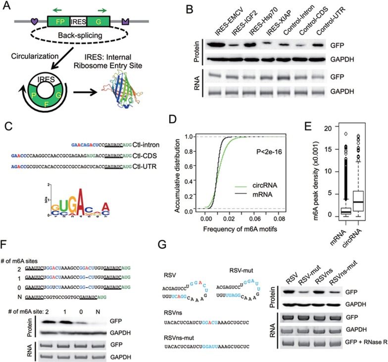
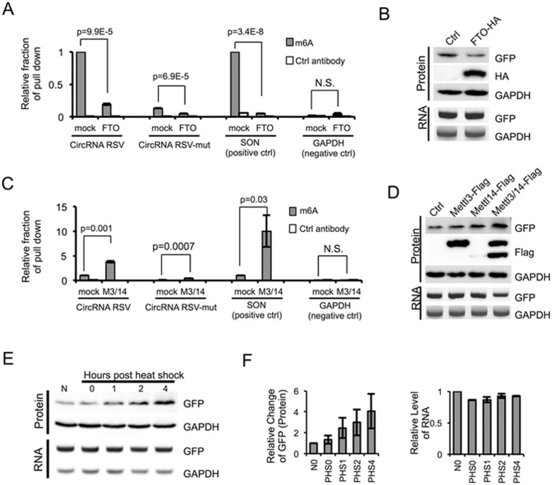
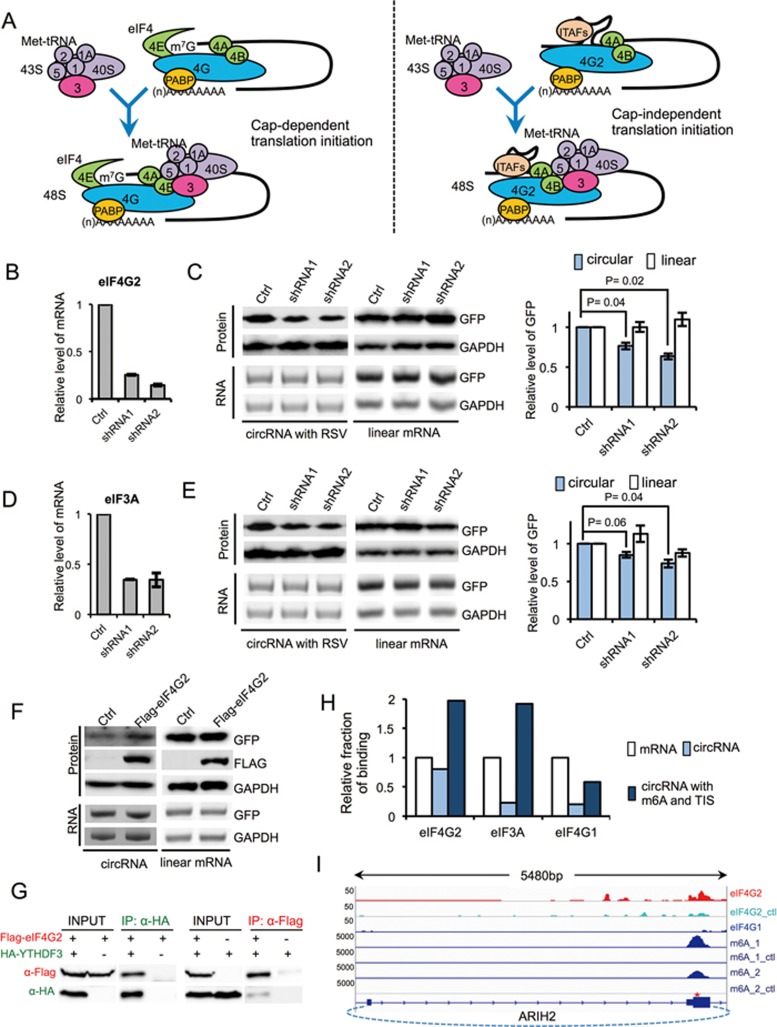
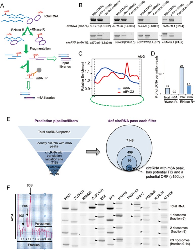
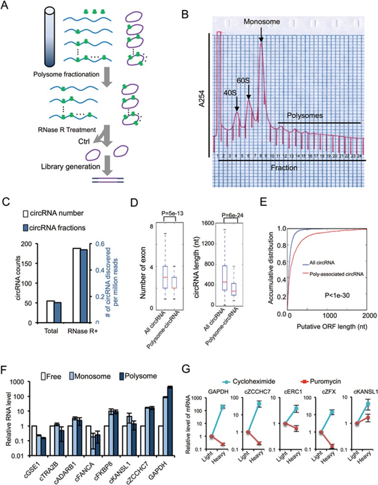
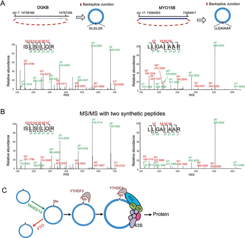
Extensive pre-mRNA back-splicing generates numerous circular RNAs (circRNAs) in human transcriptome. However, the biological functions of these circRNAs remain largely unclear. Here we report that N6-methyladenosine (m6A), the most abundant base modification of RNA, promotes efficient initiation of protein translation from circRNAs in human cells. We discover that consensus m6A motifs are enriched in circRNAs and a single m6A site is sufficient to drive translation initiation. This m6A-driven translation requires initiation factor eIF4G2 and m6A reader YTHDF3, and is enhanced by methyltransferase METTL3/14, inhibited by demethylase FTO, and upregulated upon heat shock. Further analyses through polysome profiling, computational prediction and mass spectrometry reveal that m6A-driven translation of circRNAs is widespread, with hundreds of endogenous circRNAs having translation potential. Our study expands the coding landscape of human transcriptome, and suggests a role of circRNA-derived proteins in cellular responses to environmental stress.


Figures

Figure 1
Figure 1
N6-methyladenosine promotes circRNA translation. (A) Schematic diagram of a circRNA translation reporter consisting of a single exon and two introns with complement sequences (marked by heart and crown). The exon can be back-spliced to generate circRNAs that drive GFP translation from the IRES. Green arrows indicate PCR primers used in detecting circRNA. (B) Translation of circRNA can be driven by different endogenous human IRESs (from IGF2, Hsp70 and XIAP) or three control sequences (short fragments of intron, coding region and 5′ UTR from beta-Actin gene, see Supplementary information, Table S1). Each reporter was transfected into 293 cells, and protein production was detected by western blot 48 h after transfection. CircRNA was detected by semi-quantitative PCR using circRNA specific primers. (C) Consensus m6A motifs are enriched in the “negative control” sequences. Top, RRACH motif near the start codon, with the putative m6A-modified adenosine highlighted in red and the start codon labeled in green. Bottom, enriched motifs discovered by k-mer sampling/clustering. (D) Accumulative distribution of m6A motif in circRNA and mRNA. (E) Density of m6A peaks (from MeRIP-seq) that are mapped to all mRNAs and known circRNAs regions. P-value = 3.2e−7 by student's t-test. (F) m6A motifs directly promote circRNA translation. 0-2 copies of m6A motifs (GGACU) and an adenosine-free control sequence (CGGTGCCGGTGC) were inserted into the upstream of the start codon in circRNA reporters, and circRNA and GFP translation were detected similarly as in panel B. (G) Known m6A sites (RSV and RSVns) and their mutations were tested for the activity of driving translation. Experimental procedures are the same as in panel B. In last panel, the total RNA was also treated with RNase R before RT-PCR.
Figure 2
Figure 2
Methylation of circRNA affects translation efficiency. (A) m6A in circRNA is reduced by FTO. FTO expression vector was co-transfected with circRNA containing RSV or RSV-mut m6A site into 293 cells, and the RNAs from transfected cells were pulled down by m6A-specific antibody and analyzed by RT-qPCR. The SON mRNA known to contain multiple m6A sites and GAPDH mRNA containing no m6A modification were used as controls. Control antibody is anti-GAPDH antibody. The IP experiments were repeated three times, with mean and SD plotted. (B) FTO reduces circRNA translation. RNA and protein were analyzed by semi-quantitative RT-PCR and western blots using 293 cells transfected with circRNA reporter containing RSV and FTO (or mock control). (C) METTL3 and METTL14 can methylate circRNA. circRNA with RSV or RSV-mut, METTL3 and METTL14 overexpression plasmids were co-transfected into 293 cells as in A (n = 3; mean ± SD). (D) circRNA translation is increased by METTL3/14. Experimental procedures are the same as in B. (E) 293 cells transiently expressing circRNA with RSV were subjected to heat shock stress. Cells were collected at 0, 1, 2, 4 h after heat shock (1 h at 42 °C) to analyze RNA and protein expression using semi-quantitative RT-PCR and western blots. N, no heat shock. (F) Quantification of circRNA RNA and GFP protein levels in heat-shocked cells. GAPDH levels were used for normalization (n = 3, mean ± SD).
Figure 3
Figure 3
Initiation factors eIF3A and eIF4G2 affect circRNA translation. (A) Schematic diagram of cap-dependent and cap-independent translation initiation in eukaryotic cells. In cap-dependent translation, eIF4 complex recognizes m7G and recruits 43S complex to mRNA to initiate translation. In cap-independent translation, eIF4G2 directly binds to the mRNA and recruitments 43S complex to mRNA to initiate translation. (B) eIF4G2 knockdown by two different shRNAs stably expressed in 293 cells. (C) RNAi of eIF4G2 decreases circRNA translation. 293 cells stably expressing shRNAs were transfected with circRNA reporters containing RSV sequence or linear GFP reporters (pEGFP-C1). RNA and protein expression levels were analyzed by semi-quantitative RT-PCR and western blots (left). Quantification of GFP protein levels was normalized to GAPDH (right; n = 3, mean ± SD). (D) eIF3A knockdown by two different shRNAs stably expressed in 293 cells. (E) RNAi of eIF3A decreases circRNA translation. Experimental procedures are same as in C (n = 3, mean ± SD). (F) eIF4G2 overexpression increases the circRNA translation. circRNA with RSV and eIF4G2 overexpression plasmids were co-transfected into 293 cells, and the levels of proteins and circRNAs were detected with western blots and RT-PCR. (G) Expression vectors of eIF4G2 and YTDHF3 with different epitope tags were co-expressed in 293 cells, and the anti-Flag or anti-HA antibodies were used for precipitation. (H) Relative fraction of eIF4G2, eIF3A and eIF4G1-binding sites in mRNAs, circRNAs and circRNAs with m6A site and translation initiation site. (I) eIF4G2 binding site and m6A peak in circular ARIH2 RNA.
Figure 4
Figure 4
Transcriptome-wide sequencing of m6A-modified circRNAs and predictive identification of endogenous circular mRNAs. (A) Schematic diagram of circRNA-m6A-seq protocol. (B) Validation of m6A-modified circRNAs in immunoprecipitated samples using m6A antibody or control antibody. Arrows indicate predicted circRNA size in the lanes with multiple bands, input (10%) indicates 10% of total input RNAs were used for RT-PCR, % m6A indicates percentage of m6A modification in target circRNAs (m6A antibody/(10× input (10%)). (C) Positional distribution of relative density for m6A and eIF4G2 binding site in circRNAs as compared to the respective control samples. The putative start codon was used as arbitrary marker to align the plot. When multiple AUG sites are presented in the circRNAs, the AUG that generates the longest ORF is use. (D) Number of circRNA reads (i.e., back-splice junction reads) per million of total reads in circRNA-m6A-seq samples treated with or without RNase R. (E) Schematic diagram of translatable circRNA prediction pipeline. Left, computational filters sequentially applied to identify circRNAs that contain m6A site, translation initiation site (TIS) and an ORF with sufficient length. The circRNA, m6A and TIS-sequencing data are from published results (see Materials and Methods section). Right, the numbers of circRNAs passing each filters. (F) Detection of predicted circRNAs from various host genes in polysome fractions with RT-PCRs. In the lanes with multiple bands, the circRNAs with expected size are indicated with arrows. IP, immunoprecipitation.
Figure 5
Figure 5
Systematic discovery of circular mRNA in human cells. (A) Schematic diagram of polysome-bound circRNA-seq protocol. (B) Polysome fractionation of HeLa cell lysate. All Fractions were collected. Fraction 8 was marked as R1, fraction 11 was marked as R2 and fraction 13-20 were combined together and marked as R3. Total RNA from R1, R2 and R3 were isolated separately. (C) Numbers and frequencies of circRNA junction reads detected by polysome-bound circRNA seq in samples with or without RNase R treatment. (D) Comparison of the number of exons and the length between polysome-associated circRNAs and the total circRNAs. All P-values were calculated with KS-test. (E) Accumulative distribution of the length for putative ORFs in polysome-associated circRNAs and the total circRNAs. (F) Relative ratios of monosome- and polysome-bound RNAs vs unbound (free) RNAs for several circRNAs. The linear mRNA of GAPDH was used as control. (G) HeLa cells were treated with 200 μM puromycin or cycloheximide, lysed and separated by sucrose gradient centrifugation. The RNAs from light fractions (< 60S) and heavy fraction (> 2 ribosomes) were purified and used as template for real-time RT-PCR reactions. The relative levels of RNAs associated with the heavy fraction vs the light fraction were plotted for each circRNA or GAPDH mRNA.
Figure 6
Figure 6
Identification of circRNA junction-coded peptides. (A) The collision-induced dissociation (CID) MS/MS spectrum of the [M+2H]3+ ion at m/z 465.29 of the human cDGKB peptide ISLSILQR and of human cMYO15B peptide LLGAIAAR ([M+2H]2+ ion at m/z 392.75) shown as an example. Annotated b- and y-ions are listed above and below the peptide sequence marked in red and green color, respectively. (B) The CID spectrum of MS/MS for corresponding synthetic peptides match to human cDGKB peptide ISLSILQR and cMYO15B peptide LLGAIAAR were shown as confirmation for the product of circRNA translation. Annotated b- and y-ions are listed above and below the peptide sequence marked in red and green color, respectively. (C) A schematic diagram of circRNA translation driven by m6A.

References

    1. Cocquerelle C, Daubersies P, Majerus MA, Kerckaert JP, Bailleul B. Splicing with inverted order of exons occurs proximal to large introns. EMBO J 1992; 11:1095–1098. - PMC - PubMed
    1. Nigro JM, Cho KR, Fearon ER, et al. Scrambled exons. Cell 1991; 64:607–613. - PubMed
    1. Jeck WR, Sorrentino JA, Wang K, et al. Circular RNAs are abundant, conserved, and associated with ALU repeats. RNA 2013; 19:141–157. - PMC - PubMed
    1. Memczak S, Jens M, Elefsinioti A, et al. Circular RNAs are a large class of animal RNAs with regulatory potency. Nature 2013; 495:333–338. - PubMed
    1. Salzman J, Gawad C, Wang PL, Lacayo N, Brown PO. Circular RNAs are the predominant transcript isoform from hundreds of human genes in diverse cell types. PLoS One 2012; 7:e30733. - PMC - PubMed