Case Reports
. 2017 Mar 10:7:44373.
doi: 10.1038/srep44373.

Nix restores mitophagy and mitochondrial function to protect against PINK1/Parkin-related Parkinson's disease

Affiliations
Case Reports

Nix restores mitophagy and mitochondrial function to protect against PINK1/Parkin-related Parkinson's disease

Brianada Koentjoro et al. Sci Rep. .

Abstract

Therapeutic targets are needed to develop neuroprotective treatments for Parkinson's disease (PD). Mitophagy, the selective autophagic elimination of dysfunctional mitochondria, is essential for the maintenance of mitochondrial integrity and is predominantly regulated by the PINK1/Parkin-mediated pathway. Loss of function mutations in Parkin and PINK1 cause an accumulation of dysfunctional mitochondria, leading to nigral neurodegeneration and early-onset PD with a high penetrance rate. We previously identified an asymptomatic homozygous Parkin mutation carrier who had not developed PD by her eighth decade despite the loss of functional Parkin. Here we discover a putative mechanism that protects her against PD. In contrast to Parkin-related PD patient-derived cells, the asymptomatic carrier cells show preserved mitochondrial function and mitophagy which is mediated by mitochondrial receptor Nip3-like protein X (Nix). Nix-mediated mitophagy was not affected by PINK1 knockdown. Both genetic and pharmacological induction of Nix restores mitophagy in PINK1- and Parkin-related PD patient cell lines, confirming its ability to induce mitophagy in the absence of PINK1/Parkin-mediated pathway. Moreover, Nix over-expression improves mitochondrial ATP production in these patient cells. Our results demonstrate that Nix can serve as an alternative mediator of mitophagy to maintain mitochondrial turnover, identifying Nix as a promising target for neuroprotective treatment in PINK1/Parkin-related PD.


Conflict of interest statement

The authors declare no competing financial interests.

Figures

Figure 1
Figure 1. Mitochondrial function and mitophagy are preserved in fibroblasts derived from the asymptomatic carrier.
Mitochondrial function was assessed in fibroblasts derived from healthy individuals (controls, n = 2; white bars), the asymptomatic carrier (light grey bars) and an affected Parkin-related PD patient (Parkin MT1; dark grey bars). As opposed to profound impairment in Parkin MT1, mitochondrial function in the asymptomatic carrier cells was preserved as shown by (A) the maximal rate of mitochondrial ATP synthesis, (B) representative images of cells stained with JC-1 (Scale bar: 25 μm), (C) mitochondrial membrane potential (ΔψM) determined by the ratio of red and green fluorescence signals of JC-1, (D) mitochondrial respiration determined by measuring oxygen consumption rate (OCR; mean ± SE) and (E) comparison of different aspects of mitochondrial respiration (mean ± SE). (F) Live cell imaging and (G) quantification of co-localization of autophagosomes and mitochondria were performed in the fibroblasts expressing GFP-LC3 (an autophagosomal marker, green signals in the left panels) and RFP-Mito (a mitochondrial marker, red signals in the middle panels). Exposure to CCCP caused a marked increase in co-localization of GFP-LC3 and RFP-Mito (yellow puncta in the right panels) in the control and asymptomatic carrier fibroblasts, while Parkin MT1 fibroblasts showed minimal overlapping. Scale bar: 10 μm. Elimination of mitochondria through CCCP-induced mitophagy was determined by measuring (H) mitochondrial mass using citrate synthase activity and (I) mitochondrial DNA (mtDNA) content relative to nuclear DNA (nDNA). Exposure to CCCP induced significant reduction of mitochondrial mass and mtDNA content in the controls and asymptomatic carrier fibroblasts, while no change was observed in the Parkin MT1 fibroblasts. NS; not significant, *p < 0.05, **p < 0.01, ***p < 0.001 in one-way ANOVA followed by post hoc Tukey’s HSD multiple comparison test (A,C,E,G) or in two-tailed Student’s t-test (H,I).
Figure 2
Figure 2. PINK1 is dispensable in the Parkin-independent mitophagy observed in fibroblasts derived from the asymptomatic carrier.
Successful knockdown of PINK1 by siRNA in the asymptomatic carrier fibroblasts (light grey bars) resulted in >95% decrease of (A) PINK1 transcripts (48 hours post transfection) followed by (B) a marked reduction of PINK1 protein (72 hours post transfection) compared to the scramble siRNA counterpart. Knockdown of PINK1 did not affect mitophagy in the asymptomatic carrier cells as demonstrated by the significant reduction of (C) mitochondrial mass and (D) mtDNA content by CCCP. NS; not significant, *p < 0.05, **p < 0.01, ***p < 0.001 in one-way ANOVA followed by post hoc Tukey’s HSD multiple comparison test (A,C,D) or in two-tailed Student’s t-test (B).
Figure 3
Figure 3. Identification of Nix as the mediator of the Parkin-independent mitophagy observed in fibroblasts derived from the asymptomatic carrier.
(A) Immunoblotting of Nix (35 kDa) from cell lysates of the control (white bars), the asymptomatic carrier (light grey bars) and the Parkin MT1 (dark grey bars) revealed an upregulation of Nix in the asymptomatic carrier cells regardless of CCCP treatment. β-actin (42 kDa) was used as a loading control. (B) Densitometric analysis confirmed the visual observation of Nix expression levels. (C,D) Successful knockdown of Nix by siRNA in the asymptomatic carrier fibroblasts resulted in >95% decrease of (C) Nix transcripts (48 hours post transfection) followed by (D) a marked reduction of Nix protein (72 hours post transfection) compared to the scramble siRNA counterpart. (E) Knockdown of Nix abrogated CCCP-induced co-localization of GFP-LC3 and RFP-Mito in the asymptomatic carrier fibroblasts (bottom right panel), while the scramble siRNA counterpart (upper right panel) showed an increase in their co-localization indicative of mitophagy. Scale bar: 10 μm. (F) Degree of co-localisation was significantly decreased in the asymptomatic carrier fibroblasts transfected with Nix siRNA compared to the scramble siRNA counterpart. Nix siRNA, but not the scramble siRNA, blocked the CCCP-induced reduction in (G) mitochondrial mass and (H) mitochondrial DNA (mtDNA) content compared to nuclear DNA (nDNA) in the asymptomatic carrier fibroblasts (light grey bars), while either treatment did not affect CCCP-induced mitophagy in the control fibroblasts (white bars). NS; not significant, *p < 0.05, **p < 0.01, ***p < 0.001 in one-way ANOVA followed by post hoc Tukey’s HSD multiple comparison test (B,G,H) or in two-tailed Student’s t-test (C,F).
Figure 4
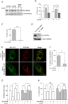
Figure 4. Nix over-expression restores mitophagy and improves mitochondrial function in fibroblasts derived from Parkin- or PINK1-related Parkinson’s disease patients.
(A) Transduction of Nix-FLAG lentivirus in fibroblasts derived from a Parkin patient (Parkin MT1) resulted in about 3-fold increase of Nix expression (48 hours post transduction) compared to the empty vector counterpart. (B) Over-expression of Nix-FLAG increased co-localization of GFP-LC3 and RFP-Mito in fibroblasts derived from the Parkin MT1 and a PINK1 patient (PINK1 MT1) upon exposure to CCCP, while the empty vector transduced counterparts showed no change. (C) The effect of Nix-FLAG on the increased co-localization in the Parkin MT1 (dark grey bars) and PINK1 MT1 (black bars) was confirmed by calculating the degree of co-localization. Exposure to CCCP induced a significant reduction in (D) mitochondrial mass and (E) mitochondrial DNA (mtDNA) content compared to nuclear DNA (nDNA) in Nix-FLAG over-expressing fibroblasts derived from Parkin (Parkin MT1, MT2 and MT3; dark grey bars) and PINK1-related Parkinson’s disease patients (PINK1 MT1 and MT2; black bars), similar to those observed in the control fibroblasts (n = 3; white bars). (F) Parkin MT1 and MT2and PINK1 MT1patient fibroblasts expressing Nix-FLAG showed a significantly higher mitochondrial ATP synthesis rate compared to the empty vector expressing counterparts. NS; not significant, *p < 0.05, **p < 0.01, ***p < 0.001 in two-tailed Student’s t-test (A) or in one-way ANOVA followed by post hoc Tukey’s HSD multiple comparison test (CF).
Figure 5
Figure 5. Pharmacological induction of Nix promotes CCCP-induced mitophagy in fibroblasts derived from Parkin or PINK1-related Parkinson’s disease patients.
(A) Treatment of phorbol 12-myristate 13-acetate (PMA; 10 nM) significantly increased Nix expression in CCCP-treated fibroblasts derived from Parkin (Parkin MT1; dark grey bars) and PINK1 (PINK1 MT1; black bars) patients. PMA-treated Parkin MT1 and PINK1 MT1 fibroblasts showed a significant reduction in (B) mitochondrial mass and (C) mitochondrial DNA (mtDNA) content compared to nuclear DNA (nDNA) upon exposure to CCCP, indicative of mitophagy, while (D and E) knockdown of Nix using siRNA, but not the scramble siRNA, abolished the PMA-mediated restoration of CCCP-induced mitophagy in both patient fibroblasts. NS; not significant, *p < 0.05, **p < 0.01 in one-way ANOVA followed by post hoc Tukey’s HSD multiple comparison test (AE).

Comment in

References

    1. Imaizumi Y. et al.. Mitochondrial dysfunction associated with increased oxidative stress and alpha-synuclein accumulation in PARK2 iPSC-derived neurons and postmortem brain tissue. Mol Brain 5, 35 (2012). - PMC - PubMed
    1. Ryan B. J., Hoek S., Fon E. A. & Wade-Martins R. Mitochondrial dysfunction and mitophagy in Parkinson’s: from familial to sporadic disease. Trends Biochem Sci 40, 200–210 (2015). - PubMed
    1. Schapira A. H. & Gegg M. Mitochondrial contribution to Parkinson’s disease pathogenesis. Parkinsons Dis 2011, 159160 (2011). - PMC - PubMed
    1. Corti O., Lesage S. & Brice A. What genetics tells us about the causes and mechanisms of Parkinson’s disease. Physiol Rev 91, 1161–1218 (2011). - PubMed
    1. Hirsch E. C., Jenner P. & Przedborski S. Pathogenesis of Parkinson’s disease. Mov Disord 28, 24–30 (2013). - PubMed

Publication types

MeSH terms