. 2017 Mar;9(1):43-52.
doi: 10.1038/ijos.2016.57. Epub 2017 Mar 10.

Distribution of pericellular matrix molecules in the temporomandibular joint and their chondroprotective effects against inflammation

Affiliations

Distribution of pericellular matrix molecules in the temporomandibular joint and their chondroprotective effects against inflammation

Wern Cui Chu et al. Int J Oral Sci. 2017 Mar.

Abstract

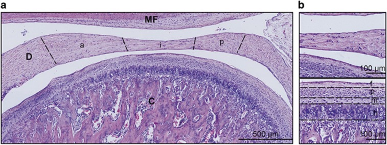
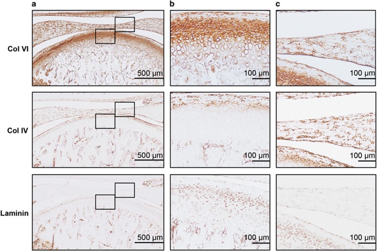
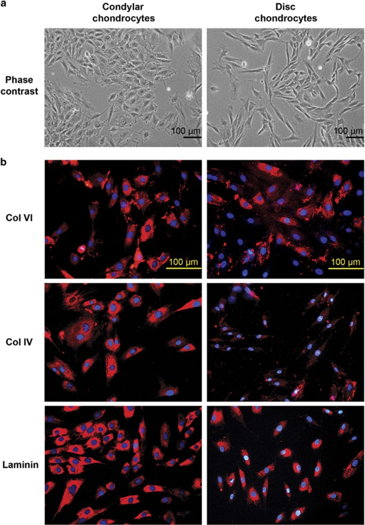
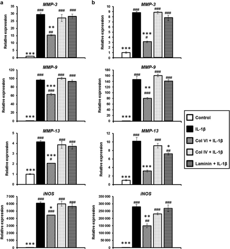
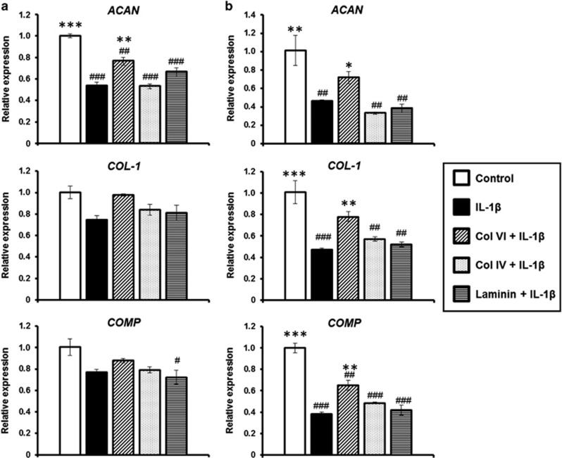
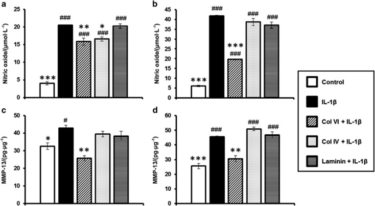
The objectives of this study were to (1) determine the distribution and synthesis of pericellular matrix (PCM) molecules (collagen VI, collagen IV and laminin) in rat temporomandibular joint (TMJ) and (2) investigate the effects of PCM molecules on chondrocytes against inflammation in osteoarthritis. Four zones (fibrous, proliferating, mature and hypertrophic) of condylar cartilage and three bands (anterior, intermediate and posterior) of disc were analysed by immunohistochemistry for the presence of PCM molecules in rat TMJs. Isolated chondrocytes were pre-treated with PCM molecules before being subjected to interleukin (IL)-1β treatment to stimulate inflammation. The responses of the chondrocytes were analysed using gene expression, nitric oxide release and matrix metalloproteinase (MMP)-13 production measures. Histomorphometric analyses revealed that the highest areal deposition of collagen VI (67.4%), collagen IV (45.7%) and laminin (52.4%) was in the proliferating zone of TMJ condylar cartilage. No significant difference in the distribution of PCM molecules was noted among the three bands of the TMJ disc. All three PCM molecules were expressed intracellularly by chondrocytes cultured in the monolayer. Among the PCM molecules, pre-treatment with collagen VI enhanced cellular proliferation, ameliorated IL-1β-induced MMP-3, MMP-9, MMP-13 and inducible nitric oxide synthase gene expression, and attenuated the downregulation of cartilage matrix genes, including collagen I, aggrecan and cartilage oligomeric matrix protein (COMP). Concurrently, collagen VI pretreatment inhibited nitric oxide and MMP-13 production. Our study demonstrates for the first time the distribution and role of PCM molecules, particularly collagen VI, in the protection of chondrocytes against inflammation.


Figures

Figure 1
Figure 1
Histological sagittal section of a rat TMJ. (a) Morphological evaluation of rat TMJ using H&E staining showing the condyle (C), disc (D), mandibular fossa (MF), and disc bands: anterior (a), intermediate (i) and posterior (p) bands. Scale bar, 500 μm. (b) Top panel shows a high magnification view of the disc. Bottom panel shows the zones of condylar cartilage: fibrous (f), proliferating (p), mature (m) and hypertrophic (h) layers. Scale bar, 100 μm. Representative images of six TMJs (n=6) harvested from three rats. TMJ, temporomandibular joint; H&E, haematoxylin and eosin.
Figure 2
Figure 2
Expression of PCM molecules in rat TMJ. (a) Histological sections of rat TMJ stained for collagen VI, collagen IV and laminin. Scale bars, 500 μm. (b) High magnification view of condylar cartilage showing the pericellular staining of collagen VI, collagen IV and laminin. Scale bars, 100 μm. (c) High magnification view of disc showing the pericellular staining of collagen VI, collagen IV and laminin. Scale bars, 100 μm. Representative images; n=6. PCM, pericellular matrix; TMJ, temporomandibular joint.
Figure 3
Figure 3
Monolayer cultures of condylar and disc chondrocytes. (a) Phase-contrast micrographs of condylar and disc chondrocytes. (b) Immunofluorescent (red) staining for collagen VI, collagen IV and laminin produced by condylar and disc chondrocytes. Nuclei were stained using DAPI (blue). Scale bars, 100 μm. Representative images; n=4 per cell type. DAPI, 4′,6-diamidino-2-phenylindole.
Figure 4
Figure 4
Effect of PCM molecules on condylar and disc chondrocyte proliferation. (a) Condylar chondrocytes and (b) disc chondrocytes treated with various concentrations of collagen VI, collagen IV or laminin were assessed for proliferation using the MTS assay at 24 and 48 h. (c) Condylar chondrocytes (left panel) and disc chondrocytes (right panel) pre-treated with collagen VI, collagen IV and laminin were subjected to IL-1β-induced inflammation for 24 h. Cell viability was assessed using the MTS assay. Mean±s.e.m.; n=4. #P<0.05; ##P<0.01; ###P<0.001, compared with untreated control. ***P<0.001, compared with IL-1β-treated group. h, hours; IL-1β, interleukin-1β PCM, pericellular matrix.
Figure 5
Figure 5
Effect of PCM molecules on matrix degradative gene expression. (a) Condylar chondrocytes and (b) disc chondrocytes pre-treated with collagen VI, collagen IV or laminin were subjected to IL-1β-induced inflammation for 6 h. Gene expression of MMP-3, MMP-9, MMP-13 and iNOS were measured using RT-PCR. Mean±s.e.m.; n=3. #P<0.05; ##P<0.01; ###P<0.001, compared with untreated control. *P<0.05, **P<0.01, ***P<0.001, compared with IL-1β-treated group. iNOS, inducible nitric oxide synthase; IL-1β, interleukin-1β MMP, matrix metalloproteinase; PCM, pericellular matrix; RT-PCR, real-time reverse transcriptase polymerase chain reaction; s.e.m.: standard error of the mean.
Figure 6
Figure 6
Effects of PCM molecules on matrix synthetic gene expression. (a) Condylar chondrocytes and (b) disc chondrocytes pre-treated with collagen VI, collagen IV or laminin were subjected to IL-1β-induced inflammation for 6 h. Gene expression of COL-1, ACAN and COMP was measured using RT-PCR. Representative results of the mean±s.e.m.; n=3. #P<0.05, ##P<0.01, ###P<0.001, compared with untreated control. *P<0.05, **P<0.01, ***P<0.001, compared with IL-1β-treated group. ACAN, aggrecan; COMP, cartilage oligomeric matrix protein; COL-1, collagen I; IL-1β, interleukin-1β PCM, pericellular matrix; RT-PCR, real-time reverse transcriptase polymerase chain reaction; s.e.m.: standard error of the mean.
Figure 7
Figure 7
Effects of PCM molecules on NO and MMP-13 production. Condylar chondrocytes (a, c) and disc chondrocytes (b, d) pre-treated with collagen VI, collagen IV or laminin were subjected to IL-1β-induced inflammation for 24 h. NO concentration in culture supernatant was measured using the Griess reaction (a, b). MMP-13 level was measured using ELISA and normalized to the total protein concentration (c,d). Mean±s.e.m.; n=3. #P<0.05, ###P<0.001, compared with untreated control. *P<0.05, **P<0.01, ***P<0.001, compared with IL-1β-treated group. ELISA, enzyme-linked immunosorbent assay; IL-1β, interleukin-1β MMP, matrix metalloproteinase; NO, nitric oxide; PCM, pericellular matrix; s.e.m.: standard error of the mean.

References

    1. Poole CA, Flint MH, Beaumont BW. Chondrons in cartilage: ultrastructural analysis of the pericellular microenvironment in adult human articular cartilages. J Orthop Res 1987; 5 (4): 509–522. - PubMed
    1. Wilusz RE, Sanchez-Adams J, Guilak F. The structure and function of the pericellular matrix of articular cartilage. Matrix Biol 2014; 39: 25–32. - PMC - PubMed
    1. Schminke B, Frese J, Bode C et al. Laminins and nidogens in the pericellular matrix of chondrocytes: their role in osteoarthritis and chondrogenic differentiation. Am J Pathol 2016; 186 (2): 410–418. - PubMed
    1. Guilak F, Alexopoulos LG, Upton ML et al. The pericellular matrix as a transducer of biomechanical and biochemical signals in articular cartilage. Ann N Y Acad Sci 2006; 1068: 498–512. - PubMed
    1. Hing WA, Sherwin AF, Poole CA. The influence of the pericellular microenvironment on the chondrocyte response to osmotic challenge. Osteoarthr Cartil 2002; 10 (4): 297–307. - PubMed

Publication types