. 2017 Apr 17;30(4):980-995.
doi: 10.1021/acs.chemrestox.6b00389. Epub 2017 Mar 29.

Mass Spectrometry Based Proteomics Study of Cisplatin-Induced DNA-Protein Cross-Linking in Human Fibrosarcoma (HT1080) Cells

Affiliations

Mass Spectrometry Based Proteomics Study of Cisplatin-Induced DNA-Protein Cross-Linking in Human Fibrosarcoma (HT1080) Cells

Xun Ming et al. Chem Res Toxicol. .

Abstract

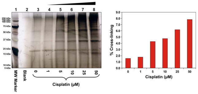
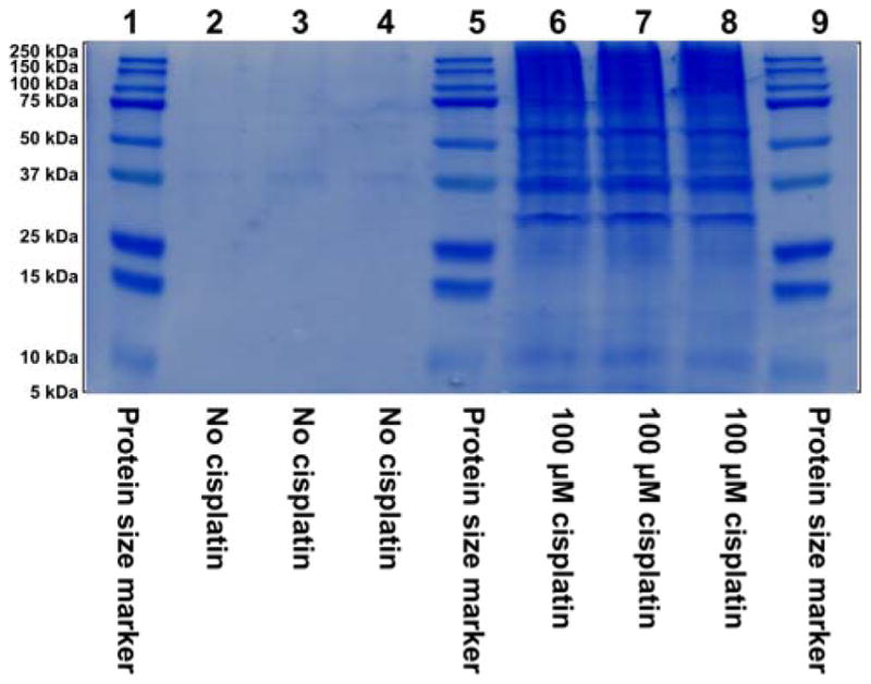
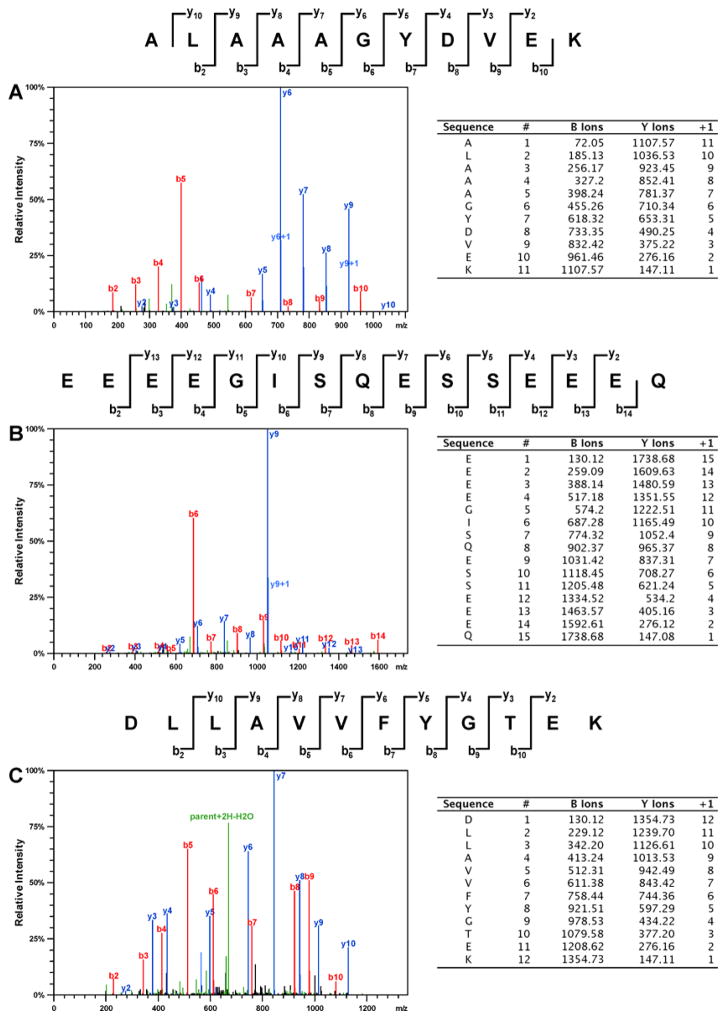
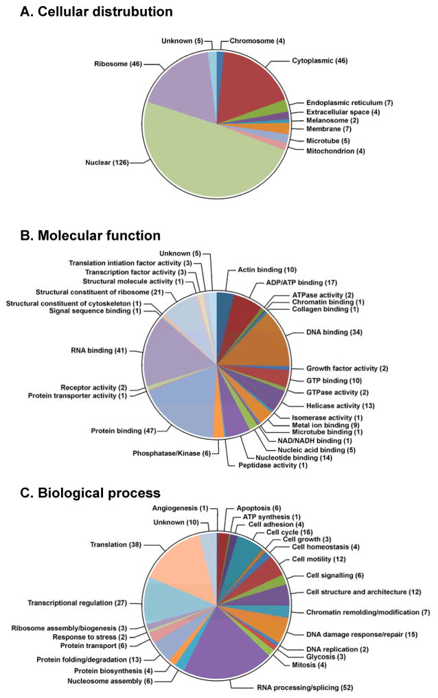
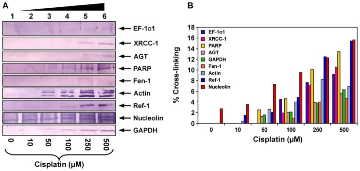
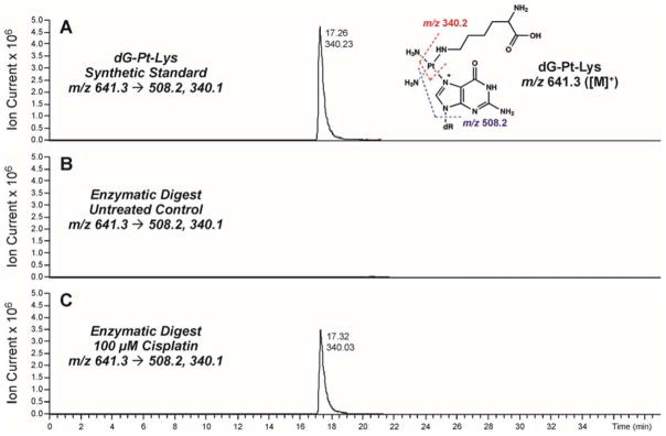
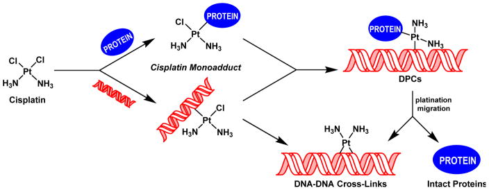
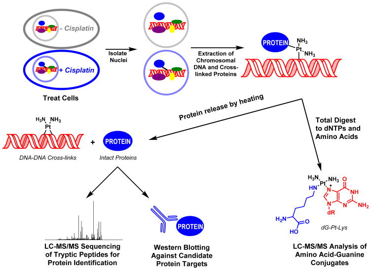
Platinum-based antitumor drugs such as 1,1,2,2-cis-diamminedichloroplatinum(II) (cisplatin), carboplatin, and oxaliplatin are currently used to treat nearly 50% of all cancer cases, and novel platinum based agents are under development. The antitumor effects of cisplatin and other platinum compounds are attributed to their ability to induce interstrand DNA-DNA cross-links, which are thought to inhibit tumor cell growth by blocking DNA replication and/or preventing transcription. However, platinum agents also induce significant numbers of unusually bulky and helix-distorting DNA-protein cross-links (DPCs), which are poorly characterized because of their unusual complexity. We and others have previously shown that DPCs block DNA replication and transcription and causes toxicity in human cells, potentially contributing to the biological effects of platinum agents. In the present work, we have undertaken a system-wide investigation of cisplatin-mediated DNA-protein cross-linking in human fibrosarcoma (HT1080) cells using mass spectrometry-based proteomics. DPCs were isolated from cisplatin-treated cells using a modified phenol/chloroform DNA extraction in the presence of protease inhibitors. Proteins were released from DNA strands and identified by mass spectrometry-based proteomics and immunological detection. Over 250 nuclear proteins captured on chromosomal DNA following treatment with cisplatin were identified, including high mobility group (HMG) proteins, histone proteins, and elongation factors. To reveal the exact molecular structures of cisplatin-mediated DPCs, isotope dilution HPLC-ESI+-MS/MS was employed to detect 1,1-cis-diammine-2-(5-amino-5-carboxypentyl)amino-2-(2'-deoxyguanosine-7-yl)-platinum(II) (dG-Pt-Lys) conjugates between the N7 guanine of DNA and the ε-amino group of lysine. Our results demonstrate that therapeutic levels of cisplatin induce a wide range of DPC lesions, which likely contribute to both target and off target effects of this clinically important drug.


Figures

Figure 1
Figure 1
Concentration-dependent formation of DPCs in nuclear protein extracts prepared from HeLa human cervical carcinoma cells following exposure to cisplatin. (A) Nuclear protein extracts from HeLa cells (500 μg) and 5′-biotinylated double-stranded oligodeoxynucleotides (3.12 nmol) were incubated in the presence of 0–50 μM Cisplatin. The resulting DPCs were captured on streptavidin beads, and the proteins were resolved on 12% SDS-PAGE. Gels were stained with SilverQuest SilverStain to visualize the cross-linked proteins. (B) Densitometric analysis of protein bands in the 25 – 250 kDa molecular weight range was used to estimate the extent of total protein cross-linking to DNA in the presence of cisplatin. Known amounts of nuclear protein extract were analyzed as a control to estimate the cross-linking efficiency.
Figure 2
Figure 2
SDS-PAGE analysis of samples employed in the proteomics studies of cisplatin-induced DPCs. HT1080 cells (~107 in triplicate) were incubated for 3 h in absence (lanes 2–4) or presence (lanes 6–8) of 100 μM cisplatin, and proteins covalently attached to chromosomal DNA were isolated as described in the Methods section. Proteins were resolved by 12% SDS-PAGE and visualized by staining with SimplyBlue SafeStatin. Molecular weight markers (lanes 1, 5 and 9) were included to permit subsequent recovery of proteins from distinct molecular weight ranges as described in the text.
Figure 3
Figure 3
Representative HPLC-ESI+-MS/MS spectra of tryptic peptides used in the identification of DPCs involving histone H1D (A), HMG B1 (B), and XRCC-6 protein (C).
Figure 4
Figure 4
GO annotations for proteins involved in cisplatin-induced DPC formation in human HT1080 cells: cellular distributions (A), molecular functions (B), and biological processes (C). The numbers of proteins in each category is indicated in parentheses.
Figure 5
Figure 5
Western blot analysis of cisplatin-induced DPCs in HT1080 cells. Following treatment with 0 (lane 1), 10 (lane 2), 50 (lane 3), 100 (lane 4), 250 (lane 5), or 500 μM cisplatin (lane 6), DNA and cross-linked proteins were isolated by phenol/chloroform extraction. Samples were normalized for DNA content, proteins from 30 μg DNA were released by thermal hydrolysis, separated by SDS-PAGE, and transferred to nitrocellulose membranes. Western blotting was performed using primary antibodies specific for EF-1α1, AGT, Fen-1, nucleolin, actin, GAPDH, PARP, Ref-1, andXRCC-1 (A). The efficiency of DPC formation in the presence of cisplatin was estimated by densitometric analysis of protein bands in DPC samples and a whole cell protein lysate control (B).
Figure 6
Figure 6
HPLC-ESI+-MS/MS analysis of dG-Pt-Lys conjugates in total proteolytic digests of chromosomal DNA recovered from cisplatin-treated cells. HT1080 cells were treated with 100 μM cisplatin for 3 h to induce DNA-protein cross-links. Following extraction of the chromosomal DNA containing covalent DPCs, the cross-linked proteins were subjected to enzymatic hydrolysis to release amino acid-nucleobase conjugates. Synthetic dG-Pt-Lys (A); enzymatic digests of DPC mixtures from HT1080 cells incubated in the absence of cisplatin (B); enzymatic digests of DPC mixtures treated with 100 μM cisplatin (C).
Scheme 1
Scheme 1
Formation of DNA-DNA cross-links and DNA-protein cross-links (DPC) by cisplatin.
Scheme 2
Scheme 2
Strategy for the isolation and analysis of DPCs from cisplatin-treated mammalian cell cultures.

References

    1. Barker S, Weinfeld M, Zheng J, Li L, Murray D. Identification of mammalian proteins cross-linked to DNA by ionizing radiation. J Biol Chem. 2005;280:33826–33838. - PubMed
    1. Zhitkovich A, Voitkun V, Kluz T, Costa M. Utilization of DNA-protein cross-links as a biomarker of chromium exposure. Environ Health Perspect. 1998;106(Suppl 4):969–974. - PMC - PubMed
    1. Thomas CB, Kohn KW, Bonner WM. Characterization of DNA-protein cross-links formed by treatment of L1210 cells and nuclei with bis(2-chloroethyl)methylamine (nitrogen mustard) Biochemistry. 1978;17:3954–3958. - PubMed
    1. Baker JM, Parish JH, Curtis JP. DNA-DNA and DNA-protein cross-linking and repair in Neurospora crassa following exposure to nitrogen mustard. Mutat Res. 1984;132:171–179. - PubMed
    1. Loeber RL, Michaelson-Richie ED, Codreanu SG, Liebler DC, Campbell CR, Tretyakova NY. Proteomic analysis of DNA-protein cross-linking by antitumor nitrogen mustards. Chem Res Toxicol. 2009;22:1151–1162. - PMC - PubMed

Publication types

MeSH terms

LinkOut - more resources