. 2017 Mar 8;18(3):583.
doi: 10.3390/ijms18030583.

A Pronounced Inflammatory Activity Characterizes the Early Fracture Healing Phase in Immunologically Restricted Patients

Affiliations

A Pronounced Inflammatory Activity Characterizes the Early Fracture Healing Phase in Immunologically Restricted Patients

Paula Hoff et al. Int J Mol Sci. .

Abstract

Immunologically restricted patients such as those with autoimmune diseases or malignancies often suffer from delayed or insufficient fracture healing. In human fracture hematomas and the surrounding bone marrow obtained from immunologically restricted patients, we analyzed the initial inflammatory phase on cellular and humoral level via flow cytometry and multiplex suspension array. Compared with controls, we demonstrated higher numbers of immune cells like monocytes/macrophages, natural killer T (NKT) cells, and activated T helper cells within the fracture hematomas and/or the surrounding bone marrow. Also, several pro-inflammatory cytokines such as Interleukin (IL)-6 and Tumor necrosis factor α (TNFα), chemokines (e.g., Eotaxin and RANTES), pro-angiogenic factors (e.g., IL-8 and Macrophage migration inhibitory factor: MIF), and regulatory cytokines (e.g., IL-10) were found at higher levels within the fracture hematomas and/or the surrounding bone marrow of immunologically restricted patients when compared to controls. We conclude here that the inflammatory activity on cellular and humoral levels at fracture sites of immunologically restricted patients considerably exceeds that of control patients. The initial inflammatory phase profoundly differs between these patient groups and is probably one of the reasons for prolonged or insufficient fracture healing often occurring within immunologically restricted patients.

Keywords: bone healing; chemokines; cytokines; fracture healing; fracture hematoma; immune cells; immunologically restricted patients; inflammation.


Conflict of interest statement

The authors declare no conflict of interest.

Figures

Figure 1
Figure 1
Monocytes/macrophages, hematopoietic stem and progenitor cells, natural killer T (NKT) cells and activated T helper cells accumulate in fracture hematoma (FH)/surrounding bone marrow (SBM) of immunologically restricted (FH IR/SBM IR) patients while regulatory T cells are decreased in SBM. Leukocytes from FH IR/SBM IR of immunologically restricted patients (dark grey) and controls (light grey) were isolated, stained with various surface markers and analyzed by flow cytometry. (A) Within all leukocytes, the CD14+ monocytes/macrophages were detected; (B) CD34+ hematopoietic stem and progenitor cells were detected within the mononuclear cells; (C) CD3+CD56+ NKT cells were detected within lymphocytes; (D) The activated CD45RA−CD25+CD3+CD4+ T helper cells were detected within the whole T helper cell population; (E) CD25+CD127−CD3+CD4+ regulatory T cells were detected within the whole T helper cell population. Controls n = 42, IR patients n = 20. IR-patients vs. controls Mann-Whitney U test, FH vs. corresponding SBM Wilcoxon test, p-values < 0.05 are shown as numbers in the Figure.
Figure 2
Figure 2
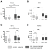
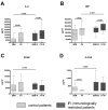
Pro-inflammatory cytokines are found at high concentrations in FH/SBM of immunologically restricted patients (FH IR/SBM IR). Supernatants from FH and SBM of immunologically restricted patients (FH IR/SBM IR: dark grey) and controls (FH/SBM: light grey) were analyzed for the concentrations of pro-inflammatory cytokines via multiplex suspension array. (A) Interleukin (IL)-1β; (B) IL-6; (C) IL-9; (D) IL-12; (E) Interferon γ (IFN)γ; (F) Tumor necrosis factor α (TNFα). Controls n = 42, IR patients n = 20. IR-patients vs. controls Mann-Whitney U test, FH vs. corresponding SBM Wilcoxon test, p-values < 0.05 are shown as numbers in the Figure.
Figure 3
Figure 3
Regulatory cytokines are increased in FH/SBM of immunologically restricted patients. Supernatants from FH and SBM of immunologically restricted patients (FH IR/SBM IR: dark grey) and controls (FH/SBM: light grey) were analyzed for the concentrations of regulatory cytokines via multiplex suspension array. (A) IL-10; (B) IL-13. Controls n = 42, IR patients n = 20. IR-patients vs. controls Mann-Whitney U test, FH vs. corresponding SBM Wilcoxon test, p-values < 0.05 are shown as numbers in the Figure.
Figure 4
Figure 4
Chemokines are up-regulated in FH/SBM of immunologically restricted patients. Supernatants from FH and SBM of immunologically restricted patients (FH IR/SBM IR: dark grey) and controls (FH/SBM: light grey) were analyzed for the concentrations of chemokines via multiplex suspension array. (A) Eotaxin/CCL11; (B) Interferon gamma-induced protein 10 (IP-10/CXCL10); (C) Macrophage inflammatory protein 1α (MIP-1α/CCL3); (D) Regulated on activation, normal T cell expressed and secreted (RANTES/CCL5). Controls n = 42, IR patients n = 20. IR-patients vs. controls Mann-Whitney U test, FH vs. corresponding SBM Wilcoxon test, p-values < 0.05 are shown as numbers in the Figure.
Figure 5
Figure 5
Angiogenic factors are highly secreted in FH/SBM of immunologically restricted patients. Supernatants from FH and SBM of immunologically restricted patients (FH IR/SBM IR: dark grey) and controls (FH/SBM: light grey) were analyzed for the concentrations of angiogenic factors via multiplex suspension array. (A) IL-8; (B) Macrophage migration inhibitory factor (MIF); (C) Platelet-derived growth factor (PDGF); (D) Granulocyte-colony stimulating factor (G-CSF). Controls n = 42, IR patients n = 20. IR-patients vs. controls Mann-Whitney U test, FH vs. corresponding SBM Wilcoxon test, p-values < 0.05 are shown as numbers in the Figure.
Figure 6
Figure 6
Summary of differences in FH/SBM of immunologically restricted patients when compared to control patients, and the influence on initial bone healing (green arrows connect suggested main sources to cytokines released; red arrows connect released cytokines to their suggested main target mechanisms and mode of action—↑up-regulation/↓down-regulation—in bone healing).

References

    1. Chidrawar S.M., Khan N., Chan Y.L., Nayak L., Moss P.A. Ageing is associated with a decline in peripheral blood CD56bright NK cells. Immun. Ageing. 2006;3:10. doi: 10.1186/1742-4933-3-10. - DOI - PMC - PubMed
    1. Ishikawa M., Nishioka M., Hanaki N., Miyauchi T., Kashiwagi Y., Kawasaki Y., Miki H., Kagawa H., Ioki H., Nakamura Y. Postoperative host responses in elderly patients after gastrointestinal surgery. Hepatogastroenterology. 2006;53:730–735. - PubMed
    1. Kang S.C., Matsutani T., Choudhry M.A., Schwacha M.G., Rue L.W., Bland K.I., Chaudry I.H. Are the immune responses different in middle-aged and young mice following bone fracture, tissue trauma and hemorrhage? Cytokine. 2004;26:223–230. doi: 10.1016/j.cyto.2004.03.005. - DOI - PubMed
    1. Smith R.M. Immunity, trauma and the elderly. Injury. 2007;38:1401–1404. doi: 10.1016/j.injury.2007.09.013. - DOI - PubMed
    1. Woodland D.L., Blackman M.A. Immunity and age: Living in the past? Trends Immunol. 2006;27:303–307. doi: 10.1016/j.it.2006.05.002. - DOI - PMC - PubMed

MeSH terms

LinkOut - more resources