. 2017 Mar 8;18(3):591.
doi: 10.3390/ijms18030591.

GSK3β and VDAC Involvement in ER Stress and Apoptosis Modulation during Orthotopic Liver Transplantation

Affiliations

GSK3β and VDAC Involvement in ER Stress and Apoptosis Modulation during Orthotopic Liver Transplantation

Mohamed Amine Zaouali et al. Int J Mol Sci. .

Abstract

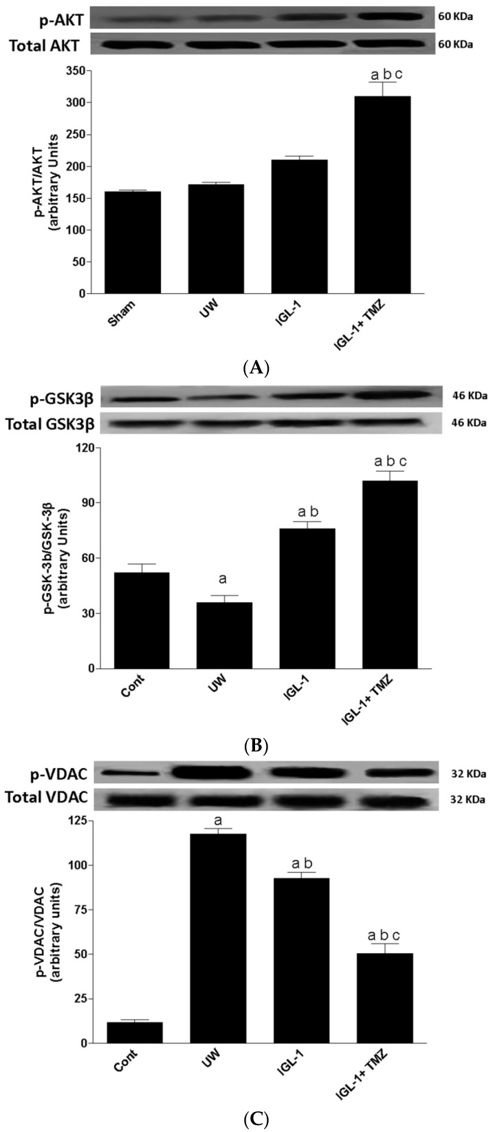
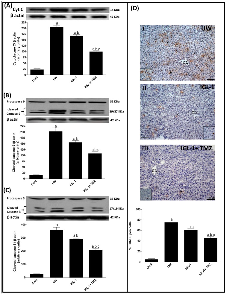
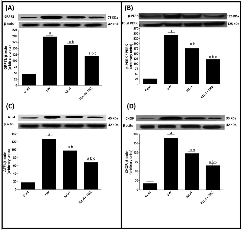
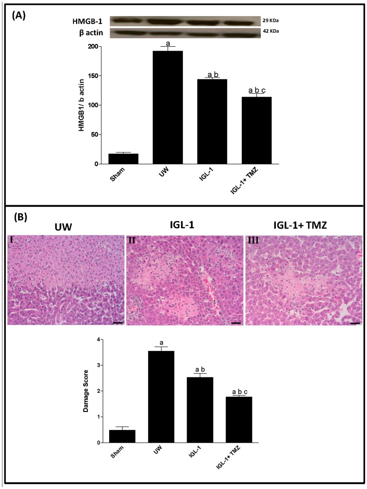
We investigated the involvement of glycogen synthase kinase-3β (GSK3β) and the voltage-dependent anion channel (VDAC) in livers subjected to cold ischemia-reperfusion injury (I/R) associated with orthotopic liver transplantation (OLT). Rat livers were preserved in University of Wisconsin (UW) and Institute Georges Lopez (IGL-1) solution, the latter enriched or not with trimetazidine, and then subjected to OLT. Transaminase (ALT) and HMGB1 protein levels, glutamate dehydrogenase (GLDH), and oxidative stress (MDA) were measured. The AKT protein kinase and its direct substrates, GSK3β and VDAC, as well as caspases 3, 9, and cytochrome C and reticulum endoplasmic stress-related proteins (GRP78, pPERK, ATF4, and CHOP), were determined by Western blot. IGL-1+TMZ significantly reduced liver injury. We also observed a significant phosphorylation of AKT, which in turn induced the phosphorylation and inhibition of GSK3β. In addition, TMZ protected the mitochondria since, in comparison with IGL-1 alone, we found reductions in VDAC phosphorylation, apoptosis, and GLDH release. All these results were correlated with decreased ER stress. Addition of TMZ to IGL-1 solution increased the tolerance of the liver graft to I/R injury through inhibition of GSK3β and VDAC, contributing to ER stress reduction and cell death prevention.

Keywords: ER stress; GSK3β; IGL-1 preservation solution; VDAC; ischemia–reperfusion injury; liver transplantation; trimetazidine.


Conflict of interest statement

The authors declare no conflict of interest.

Figures

Figure 1
Figure 1
AKT, GSK3β, and VDAC protein levels after transplantation. Representative Western blot at the top and densitometric analysis at the bottom of total and phospho-AKT (A), total and phospho-GSK3β (B) and phospho-VDAC (C). UW: liver preserved in UW solution; IGL-1: liver preserved in IGL-1 solution; IGL-1+TMZ: liver preserved in IGL-1 solution with trimetazidine. a p < 0.05 vs. Sham, b p < 0.05 vs. UW, and c p < 0.05 vs. IGL-1.
Figure 2
Figure 2
Cell death after liver transplantation. Representative Western blot at the top and densitometric analysis at the bottom of protein levels of cytochrome C (A), cleaved caspase 9 (B), and cleaved caspase 3 (C) expression in liver grafts; and (D) Representative light photomicrographs of Terminal deoxynucleotidyl transferase dUTP nick end labeling (TUNEL) stained sections. Livers submitted to Orthotopic liver transplantation (OLT) after cold storage preservation with UW (I) showed numerous positive cells, both hepatocytes (arrows) and sinusoidal lining cells (arrow heads). The positivity decreased when the livers were submitted to OLT after cold storage preservation with IGL-1 solution (II); The addition of TMZ to IGL-1 solution (III) induced a further reduction of TUNEL positivity both for hepatocytes and sinusoidal lining cells. UW: liver preserved in UW solution; IGL-1: liver preserved in IGL-1 solution; IGL-1+TMZ: liver preserved in IGL-1 solution with trimetazidine. a p < 0.05 vs. Sham; b p < 0.05 vs. UW; and c p < 0.05 vs. IGL-1. CL: centrolobular vein. Scale bar (black) indicates 50 μm.
Figure 3
Figure 3
Endoplasmic reticulum stress after liver transplantation. Representative Western blot at the top and densitometric analysis at the bottom of (A) GRP78, (B) total and phospho-PERK, (C) ATF4 and (D) Chop. UW: liver preserved in UW solution; IGL-1: liver preserved in IGL-1 solution; IGL-1+TMZ: liver preserved in IGL-1 solution with trimetazidine. a p < 0.05 vs. Sham; b p < 0.05 vs. UW; and c p < 0.05 vs. IGL-1.
Figure 4
Figure 4
Representative light photomicrographs of HSP70 (A) and HO-1 (B) immunohistochemistry. For HSP70 (A) the positivity occurred in parenchymal cells, while non-parenchymal cells displayed practically no staining, and were mainly distributed in the periportal areas. Only a few scattered hepatocytes were positive to HSP70 staining in livers submitted to OLT after cold storage preservation with UW (I); the positivity increased when the livers were submitted to OLT after cold storage with IGL-1 solution (II), and even more when trimetazidine (IGL-1+TMZ) was added to IGL-1 solution (III). Concerning HO-1, (B) the positivity occurred both in parenchymal cells (arrows) and in sinusoidal lining cells (arrow heads) and it was homogeneously distributed throughout the lobule. Livers submitted to OLT after cold storage preservation with UW (I) showed only scarce positivity. The positivity increased when the livers were submitted to OLT after cold storage preservation with IGL-1 solution (II). The addition of TMZ to IGL-1 solution (IGL1+TMZ) (III) induced a further increase of HO-1 expression both in hepatocytes and in sinusoidal lining cells. CL: centrolobular vein; P: portal vein. Scale bar 50 μm.
Figure 5
Figure 5
Inflammatory response and histological lesions in the liver. (A) Representative Western blot at the top and densitometric analysis at the bottom of HMBG1 protein levels; (B) Histological damage after liver transplantation from grafts preserved in UW (I), IGL-1 (II) and IGL-1+TMZ (III) preservation solutions. (I) Liver preserved in UW solution: extensive and confluent areas of coagulative hepatic necrosis with neutrophil infiltration; (II) Liver preserved in IGL-1 solution: reduced areas of coagulative hepatic necrosis with neutrophil infiltration; and (III) Liver preserved in IGL-1+IGL-1 solution: TMZ induced further reductions in areas of coagulative hepatic necrosis with neutrophil infiltration. UW: liver preserved in UW solution; IGL-1: liver preserved in IGL-1 solution; IGL-1+TMZ: liver preserved in IGL-1 solution with trimetazidine. Scale bar (Black) indicates 50 μm. a p < 0.05 vs. sham; b p < 0.05 vs. UW; and c p < 0.05 vs. IGL-1.

Similar articles

Cited by

References

    1. Zhai Y., Petrowsky H., Hong J.C., Busuttil R.W., Kupiec-Weglinski J.W. Ischaemia-reperfusion injury in liver transplantation—From bench to bedside. Nat. Rev. Gastroenterol. Hepatol. 2013;10:79–89. doi: 10.1038/nrgastro.2012.225. - DOI - PMC - PubMed
    1. Adam R., Delvart V., Karam V., Ducerf C., Navarro F., Letoublon C., Belghiti J., Pezet D., Castaing D., le Treut Y.P., et al. Compared efficacy of preservation solutions in liver transplantation: A long-term graft outcome study from the European Liver Transplant Registry. Am. J. Transplant. 2015;15:395–406. doi: 10.1111/ajt.13060. - DOI - PubMed
    1. Bejaoui M., Pantazi E., Folch-Puy E., Baptista P.M., Garcia-Gil A., Adam R., Rosello-Catafau J. Emerging concepts in liver graft preservation. World J. Gastroenterol. 2015;21:396–407. doi: 10.3748/wjg.v21.i2.396. - DOI - PMC - PubMed
    1. Mosbah I.B., Zaouali M.A., Martel C., Bjaoui M., Abdennebi H.B., Hotter G., Brenner C., Rosello-Catafau J. IGL-1 solution reduces endoplasmic reticulum stress and apoptosis in rat liver transplantation. Cell Death Dis. 2012;3:e279. doi: 10.1038/cddis.2012.12. - DOI - PMC - PubMed
    1. Ben Mosbah I., Casillas-Ramirez A., Xaus C., Serafin A., Rosello-Catafau J., Peralta C. Trimetazidine: Is it a promising drug for use in steatotic grafts? World J. Gastroenterol. 2006;12:908–914. doi: 10.3748/wjg.v12.i6.908. - DOI - PMC - PubMed

MeSH terms

Substances