

ProNGF, but Not NGF, Switches from Neurotrophic to Apoptotic Activity in Response to Reductions in TrkA Receptor Levels
- PMID: 28282920
- PMCID: PMC5372615
- DOI: 10.3390/ijms18030599
ProNGF, but Not NGF, Switches from Neurotrophic to Apoptotic Activity in Response to Reductions in TrkA Receptor Levels
Abstract
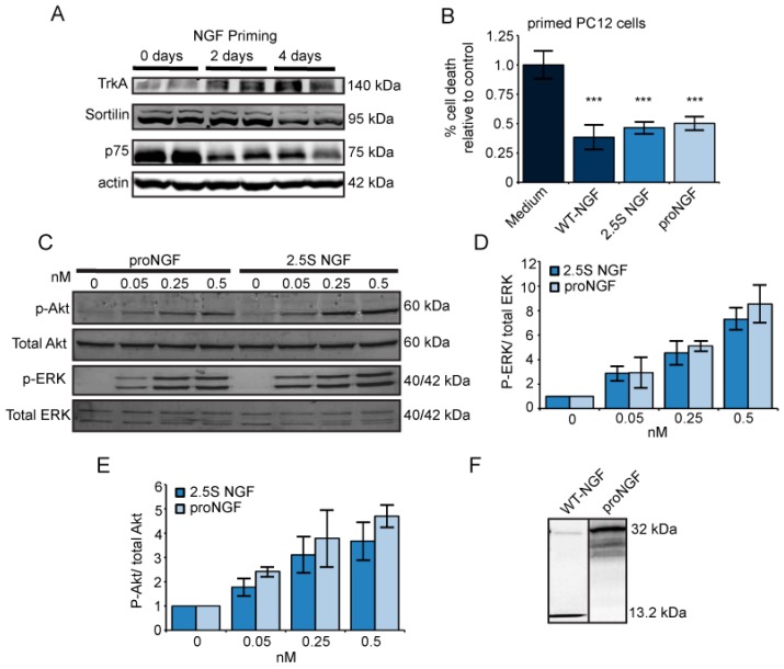
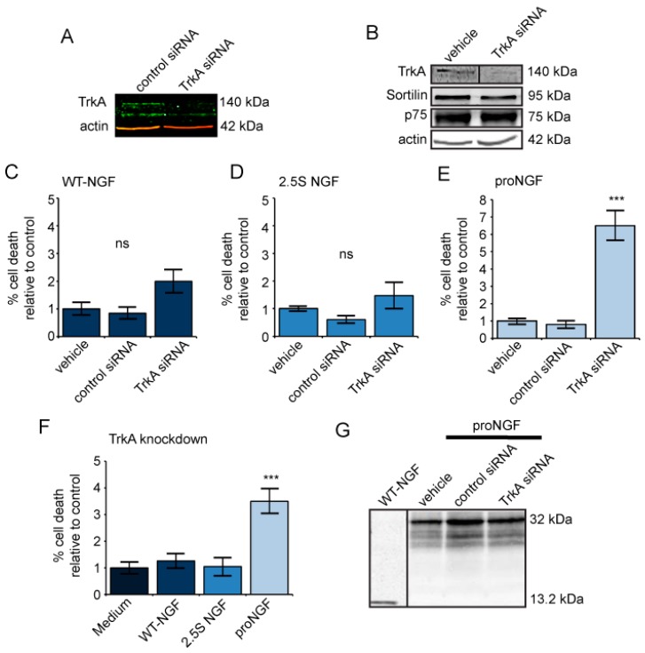
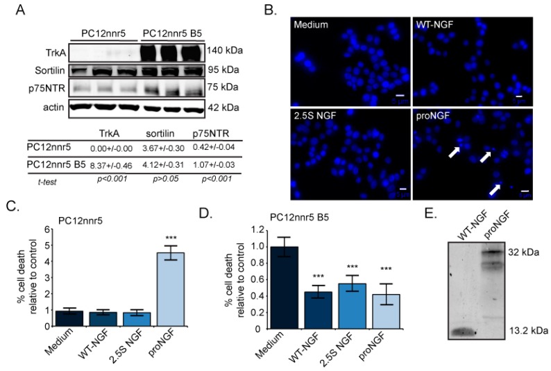
Nerve growth factor (NGF) promotes the survival and differentiation of neurons. NGF is initially synthesized as a precursor, proNGF, which is the predominant form in the central nervous system. NGF and proNGF bind to TrkA/p75NTR to mediate cell survival and to sortilin/p75NTR to promote apoptosis. The ratio of TrkA to p75NTR affects whether proNGF and mature NGF signal cell survival or apoptosis. The purpose of this study was to determine whether the loss of TrkA influences p75NTR or sortilin expression levels, and to establish whether proNGF and mature NGF have a similar ability to switch between cell survival and cell death. We systematically altered TrkA receptor levels by priming cells with NGF, using small interfering RNA, and using the mutagenized PC12nnr5 cell line. We found that both NGF and proNGF can support cell survival in cells expressing TrkA, even in the presence of p75NTR and sortilin. However, when TrkA is reduced, proNGF signals cell death, while NGF exhibits no activity. In the absence of TrkA, proNGF-induced cell death occurs, even when p75NTR and sortilin levels are reduced. These results show that proNGF can switch between neurotrophic and apoptotic activity in response to changes in TrkA receptor levels, whereas mature NGF cannot. These results also support the model that proNGF is neurotrophic under normal circumstances, but that a loss in TrkA in the presence of p75NTR and sortilin, as occurs in neurodegenerative disease or injury, shifts proNGF, but not NGF, signalling from cell survival to cell death.
Keywords: PC12 cells; TrkA; apoptosis; neurotrophin; p75NTR; proNGF; sortilin.
Conflict of interest statement
The authors declare no conflict of interest.
Figures
Similar articles
-
Type I interferons up-regulate the expression and signalling of p75 NTR/TrkA receptor complex in differentiated human SH-SY5Y neuroblastoma cells.Neuropharmacology. 2014 Apr;79:321-34. doi: 10.1016/j.neuropharm.2013.12.002. Epub 2013 Dec 11. Neuropharmacology. 2014. PMID: 24333329
-
NGF and ProNGF: Regulation of neuronal and neoplastic responses through receptor signaling.Adv Biol Regul. 2015 May;58:16-27. doi: 10.1016/j.jbior.2014.11.003. Epub 2014 Nov 20. Adv Biol Regul. 2015. PMID: 25491371 Free PMC article. Review.
-
Human ProNGF: biological effects and binding profiles at TrkA, P75NTR and sortilin.J Neurochem. 2008 Nov;107(4):1124-35. doi: 10.1111/j.1471-4159.2008.05698.x. Epub 2008 Sep 21. J Neurochem. 2008. PMID: 18808449
-
Sortilin is essential for proNGF-induced neuronal cell death.Nature. 2004 Feb 26;427(6977):843-8. doi: 10.1038/nature02319. Nature. 2004. PMID: 14985763
-
The proNGF-p75NTR-sortilin signalling complex as new target for the therapeutic treatment of Parkinson's disease.CNS Neurol Disord Drug Targets. 2008 Dec;7(6):512-23. doi: 10.2174/187152708787122923. CNS Neurol Disord Drug Targets. 2008. PMID: 19128208 Review.
Cited by
-
Urinary Levels of miR-491-5p and miR-592 as Potential Diagnostic Biomarkers in Female Aging Patients with OAB: A Pilot Study.Metabolites. 2022 Aug 31;12(9):820. doi: 10.3390/metabo12090820. Metabolites. 2022. PMID: 36144224 Free PMC article.
-
Topical delivery of nerve growth factor for treatment of ocular and brain disorders.Neural Regen Res. 2021 Sep;16(9):1740-1750. doi: 10.4103/1673-5374.306062. Neural Regen Res. 2021. PMID: 33510063 Free PMC article. Review.
-
Molecular signatures in prion disease: altered death receptor pathways in a mouse model.J Transl Med. 2024 May 27;22(1):503. doi: 10.1186/s12967-024-05121-x. J Transl Med. 2024. PMID: 38802941 Free PMC article.
-
Exploring CNS Involvement in Pain Insensitivity in Hereditary Sensory and Autonomic Neuropathy Type 4: Insights from Tc-99m ECD SPECT Imaging.Tomography. 2023 Dec 18;9(6):2261-2269. doi: 10.3390/tomography9060175. Tomography. 2023. PMID: 38133079 Free PMC article.
-
Elevated Levels of miR-144-3p Induce Cholinergic Degeneration by Impairing the Maturation of NGF in Alzheimer's Disease.Front Cell Dev Biol. 2021 Apr 9;9:667412. doi: 10.3389/fcell.2021.667412. eCollection 2021. Front Cell Dev Biol. 2021. PMID: 33898468 Free PMC article.
References
MeSH terms
Substances
LinkOut - more resources
Full Text Sources
Other Literature Sources
