. 2017 Mar 9;9(3):255.
doi: 10.3390/nu9030255.

Effects of Resveratrol Supplementation on Methotrexate Chemotherapy-Induced Bone Loss

Affiliations

Effects of Resveratrol Supplementation on Methotrexate Chemotherapy-Induced Bone Loss

Alice M C Lee et al. Nutrients. .

Abstract

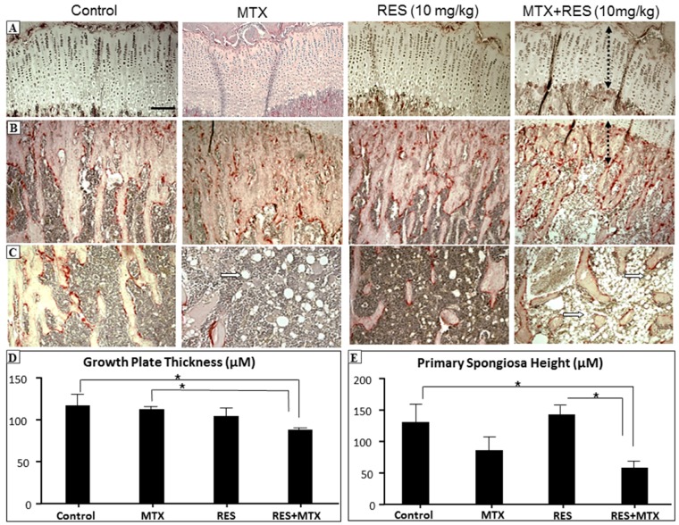
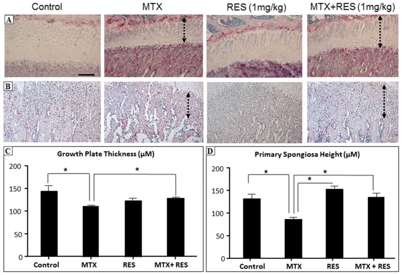
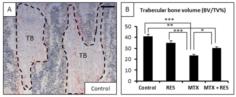
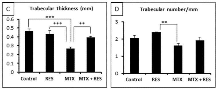
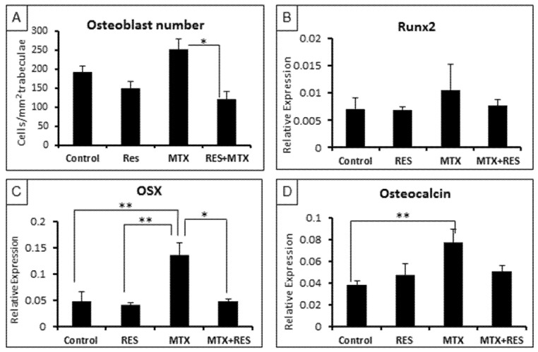
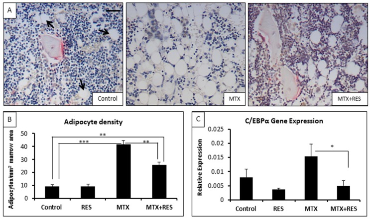
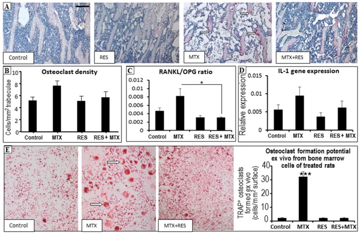
Intensive cancer chemotherapy is known to cause bone defects, which currently lack treatments. This study investigated the effects of polyphenol resveratrol (RES) in preventing bone defects in rats caused by methotrexate (MTX), a commonly used antimetabolite in childhood oncology. Young rats received five daily MTX injections at 0.75 mg/kg/day. RES was orally gavaged daily for seven days prior to, and during, five-day MTX administration. MTX reduced growth plate thickness, primary spongiosa height, trabecular bone volume, increased marrow adipocyte density, and increased mRNA expression of the osteogenic, adipogenic, and osteoclastogenic factors in the tibial bone. RES at 10 mg/kg was found not to affect bone health in normal rats, but to aggravate the bone damage in MTX-treated rats. However, RES supplementation at 1 mg/kg preserved the growth plate, primary spongiosa, bone volume, and lowered the adipocyte density. It maintained expression of genes involved in osteogenesis and decreased expression of adipogenic and osteoclastogenic factors. RES suppressed osteoclast formation ex vivo of bone marrow cells from the treated rats. These data suggest that MTX can enhance osteoclast and adipocyte formation and cause bone loss, and that RES supplementation at 1 mg/kg may potentially prevent these bone defects.

Keywords: resveratrol; bone growth arrest; adipocytes; bone loss; bone marrow adiposity; cancer chemotherapy; growth plate; methotrexate; osteoblasts; osteoclasts.


Conflict of interest statement

The authors declare no conflict of interest.

Figures

Figure 1
Figure 1
The effects of acute MTX treatment with or without resveratrol (RES) supplementation at 10 mg/kg dosage on growth plate and primary spongiosa heights and bone marrow fat content in the tibia of young rats. H and E and tartarate-resistant acidic phosphotase (TRAP) staining images showing effects of four different treatments on (A) growth plate thickness (bar = 50 μm, which applies to other images); (B) primary spongiosa heights (dashed lines); (C) bone marrow adipocytes (arrows) in the lower secondary spongiosa; (D) measurements of growth plate thickness; and (E) measurements of primary spongiosa heights. * p < 0.05.
Figure 2
Figure 2
The effects of acute MTX treatment with or without resveratrol (RES) supplementation at 1 mg/kg dosage on growth plate and primary spongiosa heights in the tibia of young rats. H and E and tartarate-resistant acidic phosphotase (TRAP) staining images showing effects of four different treatments on (A) growth plate thickness (bar = 50 μm, which applies to other images) and (B) primary spongiosa heights (dashed lines); (C) Measurements of growth plate thickness; and (D) measurements of primary spongiosa heights. * p < 0.05.
Figure 3
Figure 3
Effects of acute MTX treatment with and without resveratrol (RES) supplementation (1 mg/kg) on the trabecular bone volume and structure at the metaphysis secondary spongiosa of tibia of young rats. (A) A histological image showing traced trabecular bone (TB) of a control rat (bar = 125 μm); (B) effects on trabecular bone volume fraction BV/TV; (C) effects on trabecular thickness; and (D) effects on trabecular number. * p < 0.05, ** p < 0.01, *** p < 0.001.
Figure 3
Figure 3
Effects of acute MTX treatment with and without resveratrol (RES) supplementation (1 mg/kg) on the trabecular bone volume and structure at the metaphysis secondary spongiosa of tibia of young rats. (A) A histological image showing traced trabecular bone (TB) of a control rat (bar = 125 μm); (B) effects on trabecular bone volume fraction BV/TV; (C) effects on trabecular thickness; and (D) effects on trabecular number. * p < 0.05, ** p < 0.01, *** p < 0.001.
Figure 4
Figure 4
Effects of acute MTX treatment with and without resveratrol (RES) supplementation (1 mg/kg) on the osteoblast density and mRNA expression of osteogenesis-related genes at the metaphysis of tibia of young rats. (A) Osteoblast density on trabecular bone surface; (B) effects on expression of Runx2; (C) effects on expression of OSX; and (D) effects on expression of osteocalcin. * p < 0.05, ** p < 0.01.
Figure 5
Figure 5
Effects of acute MTX treatment with and without resveratrol (RES) supplementation (1 mg/kg) on the adipocyte density at the bone marrow of metaphysis lower secondary spongiosa of tibia of young rats. (A) Representative histology images of various groups at the lower secondary spongiosa (bar = 25 μm); (B) effects on adipocyte cell density in the bone marrow; and (C) effects on mRNA expression of adipogenesis transcription factor c/EBPα. * p < 0.05, ** p < 0.01, *** p < 0.001.
Figure 6
Figure 6
Effects of acute MTX treatment with and without resveratrol (RES) supplementation (1 mg/kg) on the osteoclast density and osteoclast formation potential in tibia of young rats. (A) Representative histology images (red colour TRAP staining as osteoclasts as indicated by arrows) of various groups at the secondary spongiosa (bar = 250 μm); (B) effects on osteoclast density on trabecular bone surface at the secondary spongiosa; (C) effects on mRNA expression ratio of osteoclastogenic factor RANKL and inhibitor OPG; (D) effects on mRNA expression of pro-osteoclastogenic cytokine IL-1; and (E) effects on osteoclast formation potentials (formed multinuclear TRAP-stained osteoclasts as indicated by arrows) of bone marrow cells from rats of various treatment groups. * p < 0.05, *** p < 0.001.

Similar articles

Cited by

References

    1. Corrie P.G. Cytotoxic chemotherapy: Clinical aspects. Medicine. 2008;36:24–28. doi: 10.1016/j.mpmed.2007.10.012. - DOI
    1. Verweij J., de Jonge M.J.A. Achievements and future of chemotherapy. Eur. J. Cancer. 2000;36:1479–1487. doi: 10.1016/S0959-8049(00)00133-7. - DOI - PubMed
    1. Sridhar T., Symonds R.P. Principles of chemotherapy and radiotherapy. Obstet. Gynaecol. Reprod. Med. 2009;19:61–67. doi: 10.1016/j.ogrm.2008.11.011. - DOI
    1. Xian C.J., Cool J.C., Scherer M.A., Macsai C.E., Fan C., Covino M., Foster B.K. Cellular mechanisms for methotrexate chemotherapy-induced bone growth defects. Bone. 2007;41:842–850. doi: 10.1016/j.bone.2007.07.021. - DOI - PubMed
    1. Fan C., Cool J.C., Scherer M.A., Foster B.K., Shandala T., Tapp H., Xian C.J. Damaging effects of chronic low-dose methotrexate usage on primary bone formation in young rats and potential protective effects of folinic acid supplementary treatment. Bone. 2009;44:61–70. doi: 10.1016/j.bone.2008.09.014. - DOI - PubMed

MeSH terms

LinkOut - more resources