. 2017 Mar 9;17(3):554.
doi: 10.3390/s17030554.

A Multimodality Hybrid Gamma-Optical Camera for Intraoperative Imaging

Affiliations

A Multimodality Hybrid Gamma-Optical Camera for Intraoperative Imaging

John E Lees et al. Sensors (Basel). .

Abstract

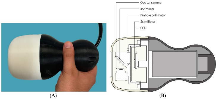
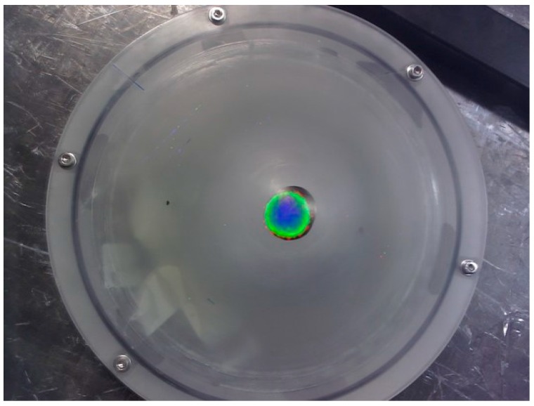
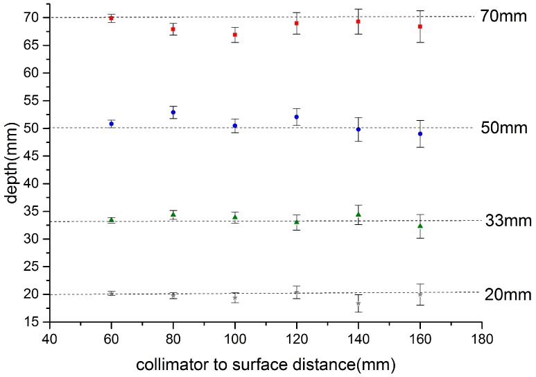
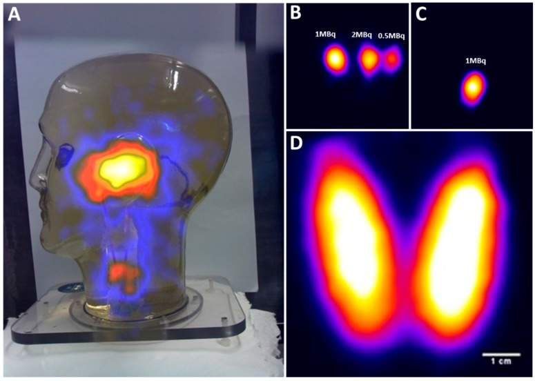
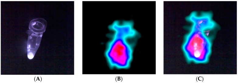
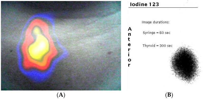
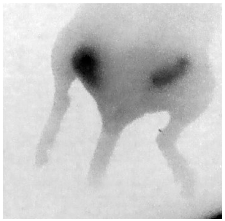
The development of low profile gamma-ray detectors has encouraged the production of small field of view (SFOV) hand-held imaging devices for use at the patient bedside and in operating theatres. Early development of these SFOV cameras was focussed on a single modality-gamma ray imaging. Recently, a hybrid system-gamma plus optical imaging-has been developed. This combination of optical and gamma cameras enables high spatial resolution multi-modal imaging, giving a superimposed scintigraphic and optical image. Hybrid imaging offers new possibilities for assisting clinicians and surgeons in localising the site of uptake in procedures such as sentinel node detection. The hybrid camera concept can be extended to a multimodal detector design which can offer stereoscopic images, depth estimation of gamma-emitting sources, and simultaneous gamma and fluorescence imaging. Recent improvements to the hybrid camera have been used to produce dual-modality images in both laboratory simulations and in the clinic. Hybrid imaging of a patient who underwent thyroid scintigraphy is reported. In addition, we present data which shows that the hybrid camera concept can be extended to estimate the position and depth of radionuclide distribution within an object and also report the first combined gamma and Near-Infrared (NIR) fluorescence images.

Keywords: NIR fluorescence; clinical images; gamma camera; hybrid imaging; optical imaging.


Conflict of interest statement

The authors declare no conflict of interest.

Figures

Figure 1
Figure 1
(A) Photograph of the Hybrid Gamma Camera (HGC); (B) schematic of the HGC showing the internal layout (for more details see Lees et al. [12]).
Figure 2
Figure 2
Schematic of the breast phantom. (A) An isometric view and; (B) a plan and side view.
Figure 3
Figure 3
Schematic of experimental arrangement of HGC and breast phantom showing a source at different heights from apex.
Figure 4
Figure 4
(A) Schematic of the insert for the head and neck phantom showing the thyroid, simplified trachea, and spine parts; (B) photograph of the complete phantom with the insert in position inside the head.
Figure 5
Figure 5
A fused optical and gamma image of a radioisotope source inside the breast phantom positioned at a distance of 70 mm below the apex.
Figure 6
Figure 6
Relationship of calculated depth versus camera-to-phantom apex point for a source at the specified set positions below the apex of the breast phantom (Figure 4). Source position: squares 70 mm, circles 50 mm, triangles 33 mm, and crosses 20 mm.
Figure 7
Figure 7
(A) A hybrid image of the head and neck phantom; (B) gamma image of three simulated parotid SLNs; (C) gamma image of simulated superficial cervical SLN; (D) gamma image of the thyroid phantom filled with 5 MBq of 99mTc activity.
Figure 8
Figure 8
(A) Image showing fluorescence dye in a small Eppendorf; (B) the gamma ray image; (C) the fused fluorescence and gamma image.
Figure 9
Figure 9
(A) Combined gamma and optical image of a patient’s thyroid during clinical investigation. The uptake of 123I is clearly seen in the right lobe of the patient’s thyroid (left side of image); (B) the standard clinical image taken by a large field of view gamma camera in the nuclear medicine clinic.
Figure 10
Figure 10
Posterior NIR fluorescence image of the hind section of a mouse showing uptake in two orthotropic tumours.

References

    1. Bluemel C., Schnelzer A., Okur A., Ehlerding A., Paepke S., Scheidhauer K., Kiechle M. Freehand SPECT for image-guided sentinel lymph node biopsy in breast cancer. Eur. J. Nucl. Med. Mol. Imaging. 2013;40:1656–1661. doi: 10.1007/s00259-013-2473-0. - DOI - PubMed
    1. Duch J. Portable gamma cameras: The real value of an additional view in the operating theatre. Eur. J. Nucl. Med. Mol. Imaging. 2011;38:633–635. doi: 10.1007/s00259-011-1761-9. - DOI - PubMed
    1. Tsuchimochi M., Hayama K. Intraoperative gamma cameras for radioguided surgery: Technical characteristics, performance parameters, and clinical applications. Phys. Med. 2013;29:126–138. doi: 10.1016/j.ejmp.2012.05.002. - DOI - PubMed
    1. Del Sordo S., Abbe L., Caroli E., Mancini A.M., Zappettini A., Ubertini P. Progress in the development of CdTe and CdZnTe semiconductor Radiation Detectors for Astrophysical and Medical Applications. Sensors. 2009;9:3491–3526. doi: 10.3390/s90503491. - DOI - PMC - PubMed
    1. Ferretti A., Chondrogiannis S., Marcolongo A., Rubello D. Phantom study of a new hand-held gamma-imaging probe for radio-guided surgery. Nucl. Med. Commun. 2013;34:86–90. doi: 10.1097/MNM.0b013e32835a7ccd. - DOI - PubMed

LinkOut - more resources