

Bacterial magnetic particles improve testes-mediated transgene efficiency in mice
- PMID: 28283003
- PMCID: PMC8241085
- DOI: 10.1080/10717544.2017.1293195
Bacterial magnetic particles improve testes-mediated transgene efficiency in mice
Abstract
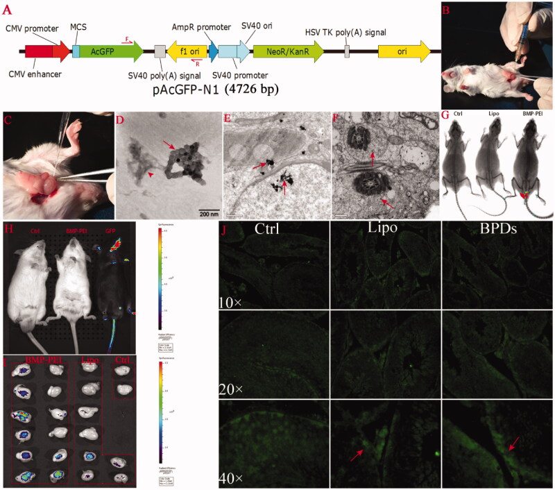
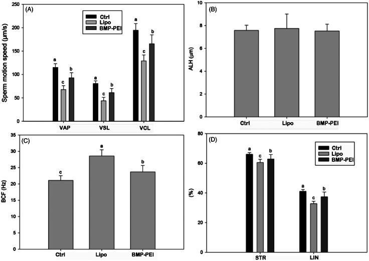
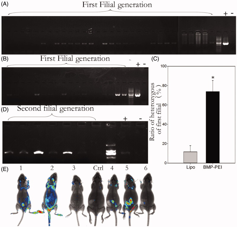
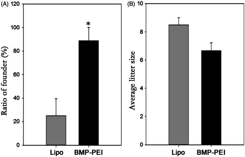
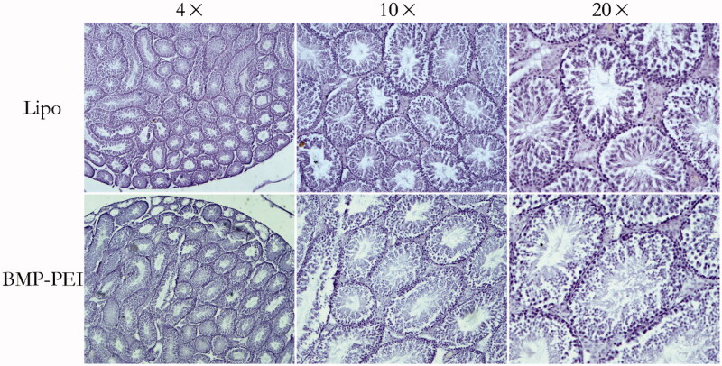
Nano-scaled materials have been proved to be ideal DNA carriers for transgene. Bacterial magnetic particles (BMPs) help to reduce the toxicity of polyethylenimine (PEI), an efficient gene-transferring agent, and assist tissue transgene ex vivo. Here, the effectiveness of the BMP-PEI complex-conjugated foreign DNAs (BPDs) in promoting testes-mediated gene transfer (TMGT) in mouse was compared with that of liposome-conjugated foreign DNAs. The results proved that through testes injection, the clusters of BPDs successfully reached the cytoplasm and the nuclear of spermatogenesis cell, and expressed in testes of transgene founder mice. Additionally, the ratio of founder mice obtained from BPDs (88%) is about 3 times higher than the control (25%) (p < 0.05). Interestingly, the motility of sperms recovered from epididymis of the founder mice from BPD group were significantly improved, as compared with the control (p < 0.01). Based on classic breeding, the ratio of transgene mice within the first filial was significantly higher in BPDs compared with the control (73.8% versus 11.6%, p < 0.05). TMGT in this study did not produce visible histological changes in the testis. In conclusion, nano-scaled BPDs could be an alternative strategy for efficiently producing transgene mice in vivo.
Keywords: Testis; bacterial magnetic particle; efficient; mice; transgene.
Conflict of interest statement
The authors report no declarations of interest.
This work was supported by National Basic Research Program of China (2013CB945501; 2014CB943202) and the Project of State Key Laboratory of Agrobiotechnology (2015SKLAB1-5).
Figures
Similar articles
-
Testis-mediated gene transfer in mice: comparison of transfection reagents regarding transgene transmission and testicular damage.Biol Res. 2011;44(3):229-34. Epub 2011 Nov 7. Biol Res. 2011. PMID: 22688909
-
Direct injection of foreign DNA into mouse testis as a possible in vivo gene transfer system via epididymal spermatozoa.Mol Reprod Dev. 2002 Jan;61(1):49-56. doi: 10.1002/mrd.1130. Mol Reprod Dev. 2002. PMID: 11774375
-
In vivo gene transfer by electroporation allows expression of a fluorescent transgene in hamster testis and epididymal sperm and has no adverse effects upon testicular integrity or sperm quality.Biol Reprod. 2006 Jan;74(1):95-101. doi: 10.1095/biolreprod.105.042267. Epub 2005 Sep 14. Biol Reprod. 2006. PMID: 16162875
-
Comparison of two methods of sperm- and testis-mediated gene transfer in production of transgenic animals: A systematic review.Anim Genet. 2024 Jun;55(3):328-343. doi: 10.1111/age.13404. Epub 2024 Feb 15. Anim Genet. 2024. PMID: 38361185
-
In vivo gene transfer into testis and sperm: developments and future application.Arch Androl. 2007 Jul-Aug;53(4):187-97. doi: 10.1080/01485010701426455. Arch Androl. 2007. PMID: 17852043 Review.
Cited by
-
Polyethylenimine-based nanocarriers in co-delivery of drug and gene: a developing horizon.Nano Rev Exp. 2018 Jul 3;9(1):1488497. doi: 10.1080/20022727.2018.1488497. eCollection 2018. Nano Rev Exp. 2018. PMID: 30410712 Free PMC article. Review.
-
Delivering CRISPR to the HIV-1 reservoirs.Front Microbiol. 2024 May 15;15:1393974. doi: 10.3389/fmicb.2024.1393974. eCollection 2024. Front Microbiol. 2024. PMID: 38812680 Free PMC article. Review.
References
-
- Amaral MG, Campos VF, Seixas FK, et al. . (2011). Testis-mediated gene transfer in mice: comparison of transfection reagents regarding transgene transmission and testicular damage. Biol Res 44:229–34. - PubMed
-
- Bian F, Mao G, Guo M, et al. . (2012). Gradients of natriuretic peptide precursor A (NPPA) in oviduct and of natriuretic peptide receptor 1 (NPR1) in spermatozoon are involved in mouse sperm chemotaxis and fertilization. J Cell Physiol 227:2230–9. - PubMed
-
- Campos VF, Komninou ER, Urtiaga G, et al. . (2011a). NanoSMGT: transfection of exogenous DNA on sex-sorted bovine sperm using nanopolymer. Theriogenology 75:1476–81. - PubMed
-
- Campos VF, Leon PM, Komninou ER, et al. . (2011b). NanoSMGT: transgene transmission into bovine embryos using halloysite clay nanotubes or nanopolymer to improve transfection efficiency. Theriogenology 76:1552–60. - PubMed
Publication types
MeSH terms
Substances
LinkOut - more resources
Full Text Sources
Other Literature Sources