. 2017 Mar 9;168(6):1015-1027.e10.
doi: 10.1016/j.cell.2017.02.019.

5' End Nicotinamide Adenine Dinucleotide Cap in Human Cells Promotes RNA Decay through DXO-Mediated deNADding

Affiliations

5' End Nicotinamide Adenine Dinucleotide Cap in Human Cells Promotes RNA Decay through DXO-Mediated deNADding

Xinfu Jiao et al. Cell. .

Abstract

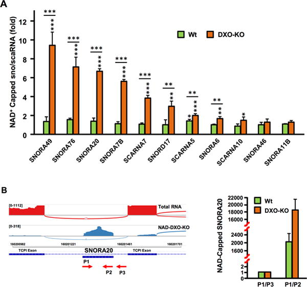
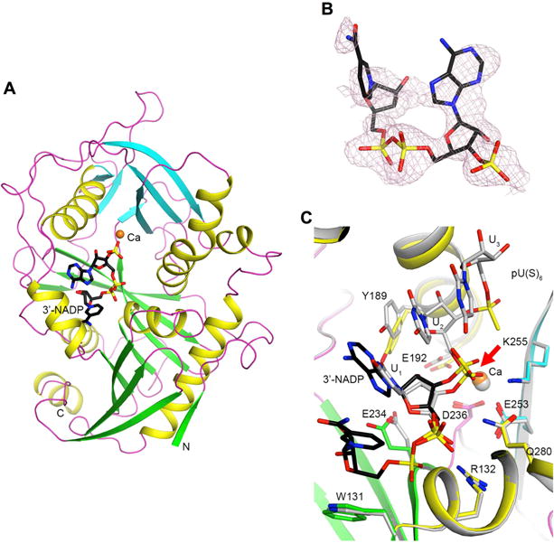
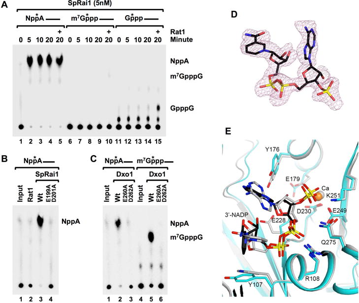
Eukaryotic mRNAs generally possess a 5' end N7 methyl guanosine (m7G) cap that promotes their translation and stability. However, mammalian mRNAs can also carry a 5' end nicotinamide adenine dinucleotide (NAD+) cap that, in contrast to the m7G cap, does not support translation but instead promotes mRNA decay. The mammalian and fungal noncanonical DXO/Rai1 decapping enzymes efficiently remove NAD+ caps, and cocrystal structures of DXO/Rai1 with 3'-NADP+ illuminate the molecular mechanism for how the "deNADding" reaction produces NAD+ and 5' phosphate RNA. Removal of DXO from cells increases NAD+-capped mRNA levels and enables detection of NAD+-capped intronic small nucleolar RNAs (snoRNAs), suggesting NAD+ caps can be added to 5'-processed termini. Our findings establish NAD+ as an alternative mammalian RNA cap and DXO as a deNADding enzyme modulating cellular levels of NAD+-capped RNAs. Collectively, these data reveal that mammalian RNAs can harbor a 5' end modification distinct from the classical m7G cap that promotes rather than inhibits RNA decay.


Figures

Figure 1
Figure 1. Mammalian cells contain NAD+-capped mRNA
(A) Selectivity of NAD-capture approach. NAD-RNA capture was carried out with 100μg HEK293T total RNA in the presence of 32P-uniform-labeled 20pm NAD+-capped or m7GpppA-capped RNA with or without ADPRC treatment as indicated. RNA bound to magnet streptavidin beads (SB) or in the flow through (FT) were isolated resolved on 8% Denaturing Urea PAGE. The asterisks represent the positions of the 32P-labeling. Quantitation of the spiked RNA from two independent experiments revealed 1% of input NAD+-capped RNA was retained on the affinity matrix under –ADPRC conditions while 89% with ADPRC. For m7G capped RNA, 1% was retained with –ADPRC while 2% was detected with ADPRC. (B) Analysis of NAD-capture RNA-Seq FPKM distribution of assembled transcript lengths by sample. Calculated transcript length was obtained from Cufflinks output. FPKM values were summarized up to the length indicated using box plots, with the center line indicating median, the top and bottom of the box indicating 75th and 25th percentiles, and the mean by the diamond-shaped symbol. Total RNA: Ribosome-minus HEK293 RNA sample processed for RNA-seq by standard methods; NAD-RNA: NAD-capture RNA prepared from HEK293 cells. (C) Sequencing reads were intersected with a general genome map (GenCode, hg19 genome). The number of features overlapping by at least 50% of length is displayed by RNA class (color key) for RNAP II transcripts. (D) Aligned sequencing reads were visualized as a Sashimi plot using the Integrative Genomics Viewer (Broad Institute) where the density of reads are denoted above LSM3 gene. Boxes on the bottom represent exons and the curved lines represent adjoining exon-exon reads. (E) The percentage of the indicated NAD+-capped mRNAs that harbor a poly(A) tail relative to its corresponding total NAD+ capped mRNA is shown. Total NAD+ capped mRNA was designated as 100. Error bars represent ±SD. (F) Sequence read of COX7B 3′ end containing 30 adenosine poly(A) tail is shown. Sequencing of 5 indiviual clones revealed a range of poly(A) tails spanning from 18 to 51 adenosines.
Figure 2
Figure 2. Mammalian DXO possesses robust deNADding activity
(A) In vitro decapping assays with 10nM mouse recombinant DXO protein and indicated 32P-cap-labeled RNA substrates. Reaction products were resolved by polyethyleneimine (PEI)-cellulose thin-layer chromatography (TLC) developed in 0.45 M (NH4)2SO4. (B) Schematic of NAD+-capped RNA and sites of DXO cleavage. The red “P” denotes the position of the 32P. (C) Quantitation from (A) carried out with ImageQuant software and plotted from three independent experiments with the error bars representing +/−SD. (D) Catalytically inactive mutants E234A and K256Q abolish DXO deNADding activity. (E) Mammalian Dcp2 does not possess intrinsic deNADding activity. Recombinant (50nM) human Dcp2, mouse DXO or bacteria NudC proteins were incubated with 32P-cap-labeled NAD-RNA at 37°C for 30 minutes. The reaction products were either untreated (−) or treated with 1U Nuclease P1 (+), resolved by PEI-TLC developed in 0.45 M (NH4)2SO4. Product standards were denoted on the right. The asterisks represent the position of the 32P-labeling. (F) Similar to B, except sites of NudC and Nuclease P1 (Nuc. P1) hydrolysis are denoted.
Figure 3
Figure 3. DXO is a deNADding enzyme in cells and NAD-capped RNAs do not support translation
(A) 32P-NAD+ cap-labeled RNA was transfected into HEK293T control knock-out (Con-KO), DXO knock-out (DXO-KO) or Dcp2 knock-out (Dcp2-KO) cell lines with Lipofectamine 3000. Untransfected RNAs were degraded with micrococcal nuclease (MN) and total RNA was isolated at the indicated time points following MN treatment and resolved on 8% Denaturing PAGE and exposed to a PhosphorImager. RNA remaining was quantitated and plotted from three independent experiments with ±SD denoted by the error bars. Stabilities: Con-KO t1/2=2.8 hr [95%CI: 2.0 – 4.5]; DXO-KO t1/2=6.3 hr [95%CI: 4.2 – 11.9] (ANOVA, p=6.10 × 10⁻⁴); Dcp2-KO t1/2=3.2 hr, [95%CI: 2.3 – 5.8] (ANOVA, p=0.38). (B) Same as A except the NAD+-capped RNA was uniformly labeled 32P. Stabilities: Con-KO t1/2=3.0 hr [95%CI: 2.4 – 4.3]; DXO-KO t1/2=5.7 hr [95%CI: 4.1 – 8.9] (ANOVA, p=7.88 × 10⁻⁴); Dcp2-KO t1/2=3.3 hr, [95%CI: 2.7 – 4.4] (ANOVA, p=0.17). (C) Same as A except the RNA contained an m7G cap and was uniformly labeled within the RNA body with 32P. Stabilities: Con-KO t1/2=4.1 hr [95%CI: 3.2 – 5.8]; DXO-KO t1/2=3.9 hr [95%CI: 3.0 – 5.4] (ANOVA, p=0.51); Dcp2-KO t1/2=4.1 hr, [95%CI: 3.1 – 5.9] (ANOVA, p=0.64). (D). Luciferase RNAs with a 5′-end triphosphate lacking a cap, 5′-end NAD+ cap or m7G cap transfected into control or DXO-KO cells. Cell were treated with micrococcal nuclease to degrade untransfected RNA, harvested at the indicated times and remaining RNA levels quantitated from three independent experiments. The left panel shows RNAs transfections into control HEK293T (Con-KO) cells and the right panel presents RNAs introduced into either Con-KO or DXO-KO cells as indicated. Error bars denoted ±SD. Con-KO m7G-FLuc t1/2= 92.6 min (95% CI: 82.2 to 107.8 min, R2=0.96, p=2.10 × 10⁻⁸); DXO-KO m7G-FLuc t1/2=125 min (95% CI: 92.2 to 174 min, R2=0.84, p=1.47 × 10⁻⁵); Con-KO NAD-FLuc t1/2= 67.4 min (95% CI: 51.4 to 97.9 min, R2=0.82, p=3.05 × 10⁻⁵); DXO-KO NAD-FLuc t1/2= 103.6 min (95% CI: 85.2 to 132.3 min, R2=0.91, p=1.22 × 10⁻⁶); Con-KO pppA-RNA=110.7 min (95% CI: 88 to 149.2 min, R2=0.870, p=5.99 × 10⁻⁵). (E). Firefly luciferase mRNAs containing either a 5′-end NAD+ cap, m7G cap or no cap and 3′ poly(A)60 tail were co-transfected with m7G-capped Renilla Luciferase RNA into HEK293T cells. Cells were harvested and assayed at the indicated time points. Firefly Luciferase activity was plotted normalized to Renilla Luciferase and data from three independent experiments are presented with error bars representing +/−SD.
Figure 4
Figure 4. DXO is a deNADding enzyme in cells
(A) Volcano plot of NAD-Capture transcripts comparing WT with DXO-KO samples. The log2 fold change (DXO-KO/WT) is plotted vs. the –log10 FDR (False Discovery Rate; corrected for multiple measurements). Each transcript is indicated as a dot on the plot. Dots above the horizontal dashed line are ≤ 5% FDR. The vertical dashed lines indicate ±2-fold differences, so dots to the right of the +2-fold line are colored black (only if they are detected with at least 1 FPKM). Red dots indicate the mRNAs assessed by qPCR in panel B and blue dots indicate snoRNAs assessed by qPCR in Fig. 5A. (B) qRT-PCR validation of NAD+ capped mRNAs in DXO-KO cells. Randomly selected NAD+-capped RNAs from the NAD-CaptureSeq were eluted from the beads, reverse transcribed and detected with gene specific primers. Data are presented relative to the -ADPRC negative control set to 1. MRPL13, which is not responsive to DXO in the NAD CaptureSeq, was included as a negative control. The inserted table represents levels of NAD+ capped RNAs relative to their respective total mRNA in the indicated cells. Error bars represent ±SD. P values are denoted by asterisks; (*) P < 0.05, (**) P < 0.01, (***) P < 0.001 (Student’s t-test).
Figure 5
Figure 5. Small nucleolar and small Cajal body RNAs can be NAD+ capped and their levels are dependent on DXO in cells
(A) A subset of NAD+ capped sno/scaRNAs were validated by qRT-PCR. NAD+-capped RNAs isolated by the NAD-Capture approach were eluted from the beads, reverse transcribed and the indicated sno/scaRNAs detected with gene specific primers. Data are presented relative to the -ADPRC negative control set to 1. SNORA46 and SNORA11B, which were not responsive to DXO in the NAD CaptureSeq, were also tested. Error bars represent ±SD. P values are denoted by asterisks; (*) P < 0.05, (**) P < 0.01, (***) P < 0.001 (Student’s t-test). (B) Sashimi plot with SNORA20 within the TCP1 intron 9 is shown. Labeling is as in Figure 1E. Detected SNORA20 reads are within the mature snoRNA. The P1 – P3 primers used to amplify SNORA20 or the SNORA20-intron junction are shown and qRT-PCR results graphed on the right.
Figure 6
Figure 6. Crystal structure of mouse DXO in complex with 3′-NADP+
(A) Schematic drawing of the structure of DXO (in green and cyan for the large and small β-sheets, respectively, yellow for helices, and magenta for loops) in complex with 3′-NADP+ (in black for carbon atoms) and one Ca2+ ion (orange). (B). Omit Fo–Fc electron density at 2.1 Å resolution for 3′-NADP+, contoured at 2σ. (C). Overlay of the binding modes of 3′-NADP+ (black) and Ca2+ (orange) with those of the pU(S)6 substrate mimic and Ca2+ (gray) (Jiao et al., 2013). The scissile phosphate is denoted with a red arrow. All the structure figures were produced with PyMOL (www.pymol.org).
Figure 7
Figure 7. SpRai1 and KlDxo1 are deNADding enzymes
(A) SpRai1 is a robust deNADding enzyme. In vitro decapping assays were carried out with 5nM recombinant SpRai1 with 32P 5′ end-cap-labeled NAD+-capped, m7GpppA-capped or GpppA-capped RNAs and products detected as in legend to Figure 3. The (+) indicates the addition of 5nM recombinant Rat1. (B) SpRai1 catalytically inactive double mutant E199A/D201A and (C) catalytically inactive Dxo1 double mutant E260A/D262A, abolish deNADding or decapping activities. (D). Omit Fo–Fc electron density at 1.9 Å resolution for 3′-NADP+ in the complex with SsRai1, contoured at 2σ. (E). Overlay of the binding modes of 3′-NADP+ (black) and Ca2+ (orange) in the complex with SsRai1 (cyan) with those in the complex with DXO (gray).

References

    1. Adams PD, Grosse-Kunstleve RW, Hung LW, Ioerger TR, McCoy AJ, Moriarty NW, Read RJ, Sacchettini JC, Sauter NK, Terwilliger TC. PHENIX: building new software for automated crystallographic structure determination. Acta Crystallogr D Biol Crystallogr. 2002;58:1948–1954. - PubMed
    1. Bird JG, Zhang Y, Tian Y, Panova N, Barvik I, Greene L, Liu M, Buckley B, Krasny L, Lee JK, et al. The mechanism of RNA 5′ capping with NAD+, NADH and desphospho-CoA. Nature. 2016;535:444–447. - PMC - PubMed
    1. Cahova H, Winz ML, Hofer K, Nubel G, Jaschke A. NAD captureSeq indicates NAD as a bacterial cap for a subset of regulatory RNAs. Nature. 2015;519:374–377. - PubMed
    1. Chang JH, Jiao X, Chiba K, Oh C, Martin CE, Kiledjian M, Tong L. Dxo1 is a new type of eukaryotic enzyme with both decapping and 5′-3′ exoribonuclease activity. Nat Struct Mol Biol. 2012;19:1011–1017. - PMC - PubMed
    1. Chen CY, Ezzeddine N, Shyu AB. Messenger RNA half-life measurements in mammalian cells. Methods Enzymol. 2008;448:335–357. - PMC - PubMed

Publication types