. 2017 Mar 9;168(6):1028-1040.e19.
doi: 10.1016/j.cell.2017.02.027.

Stress-Triggered Phase Separation Is an Adaptive, Evolutionarily Tuned Response

Affiliations

Stress-Triggered Phase Separation Is an Adaptive, Evolutionarily Tuned Response

Joshua A Riback et al. Cell. .

Abstract

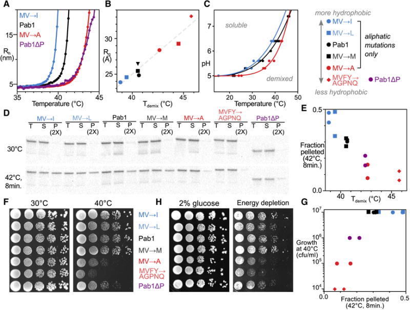
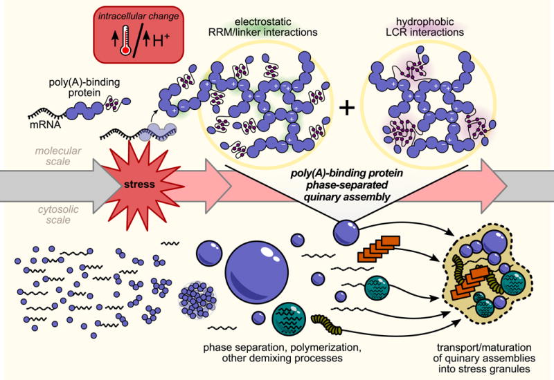
In eukaryotic cells, diverse stresses trigger coalescence of RNA-binding proteins into stress granules. In vitro, stress-granule-associated proteins can demix to form liquids, hydrogels, and other assemblies lacking fixed stoichiometry. Observing these phenomena has generally required conditions far removed from physiological stresses. We show that poly(A)-binding protein (Pab1 in yeast), a defining marker of stress granules, phase separates and forms hydrogels in vitro upon exposure to physiological stress conditions. Other RNA-binding proteins depend upon low-complexity regions (LCRs) or RNA for phase separation, whereas Pab1's LCR is not required for demixing, and RNA inhibits it. Based on unique evolutionary patterns, we create LCR mutations, which systematically tune its biophysical properties and Pab1 phase separation in vitro and in vivo. Mutations that impede phase separation reduce organism fitness during prolonged stress. Poly(A)-binding protein thus acts as a physiological stress sensor, exploiting phase separation to precisely mark stress onset, a broadly generalizable mechanism.

Keywords: RNA-binding protein; energy depletion; heat shock; intrinsically disordered protein; low-complexity region; membraneless organelle; pH; poly(A)-binding protein; quinary structure; stress granules.


Figures

Figure 1
Figure 1. Heat stress triggers formation of RNase-insensitive Pab1 quinary assemblies, separable from stress granule formation
A, Confocal fluorescent microscopy images of diploid strains, containing Pab1-mRuby2 and Pab1-Clover, showing Pab1-mRuby2 after 8-minute incubation at the indicated temperatures. Arrow indicates a stress granule. B, Pab1 western blot after 10-minute heat shock of wild-type cells. Lysed samples were incubated with or without RNase1, then progressively fractionated at 8,000g and 100,000g yielding pellets (P8 and P100, respectively) and remaining supernatant (S). C and D, Quantification of B where each fraction is normalized to the total intensity in all fractions. Red or black indicate RNase1 or buffer treatment, respectively.
Figure 2
Figure 2. Purified Pab1 demixes in response to thermal shock, releasing RNA with small changes in secondary structure
A, Top, size-exclusion chromatography trace of Pab1 after 30°C incubation (black) and after heating at 46°C for 30 min (red). Below, Pab1 with ~2:1 excess of A19 RNA treated identically. Blue trace shows A19 alone. B, Pab1 total-protein dilution for calibration (left) and pelleted material after heating with and without RNA (right), Coomassie-stained. C, DLS temperature ramp experiments of Pab1 with indicated protein to RNA ratios. D, Tdemaix at RNA concentrations from C. E, Kinetics of Pab1 assembly monitored by DLS after a temperature jump (cf. Fig. S1). F, Rate of hydration radius growth from E with the accompanying average Q1036°C value. G, Temperature jump of 0.2 μM Pab1 (top left, with numbered full-scan timepoints indicated) and accompanying CD spectra (top right). Total ellipticity between 210-250nm (bottom) shows linear temperature-independent signal attenuation used to scale scans to time zero (bottom right) (Fig. S2).
Figure 3
Figure 3. Pab1 demixing proceeds via liquid-liquid phase separation and gel formation, modulated but not caused by its low-complexity region
A, Demixing of purified Pab1 is sensitive to ionic strength and pH (Fig. S3) B, Morphology of 15 μM Pab1-mRuby2 assemblies. C, Fluorescence recovery after photobleaching of Pab1 droplets. D, Sequentially assembled two-color droplets remain unmixed after 24 hours. E, The Tdemix of Pab1 measured at different pH values defines a phase boundary. F, Pab1 domain deletions and corresponding DLS temperature ramps (Table S1). G, H, Morphology of Pab1ΔP quinary assemblies. G, 15 μM 100:3 Pab1ΔP:Pab1-Clover; H, 15 μM Pab1ΔP alone.
Figure 4
Figure 4. Evolutionary analysis reveals rapid exchange between aliphatic residues in poly(A)-binding protein’s proline-rich low-complexity region
A, The low-complexity P domain of S. cerevisiae Pab1 colored by amino acid types. B, Amino acid usage in the P domain, ordered by enrichment relative to the rest of Pab1 (Pab1ΔP). Usage for all yeast proteins, and for disordered sequences curated by DisProt, are shown for comparison. C, A diverse alignment of 295 PABPs (pruned for clarity of display from 351) indicating locations of the RRMs, P domain, and CTD where each column is a residue, colored as in A, and each row is a species. White spaces are alignment gaps. The figure contains no text. A black triangle marks a clade-specific insertion in RRM4. D, A subset of fungal species and a portion of the P domain from the alignment in C containing multiple sites where aliphatic residues (colored to show differences) exchange rapidly while nearby positions (starred) remain invariant. E, In the P domain, but not in general, aliphatic residue frequency negatively correlates with residue hydrophobicity. The mean aliphatic residue usage in the aligned set of P domains, remainder of PABP, disordered sequences from DisProt, and the yeast proteome are shown, colored as in B. Cf. Fig. S4. Error bars throughout show standard error on the mean.
Figure 5
Figure 5. The P domain is unstructured and displays hydrophobicity-dependent compaction
A, CD spectrum of the P domain (without His8 tag) at ~1μM, 20°C. Inset, the temperature dependence of the average CD signal from 215–235 nm. B, SAXS of P domain (with His8 tag) at denaturant (GdnHCl) concentrations shown at right. Inset highlights the mid-q region. Corresponding Rg values are plotted; gray line shows extrapolation to zero denaturant (see Methods). Dashed lines correspond to approximate values for denatured proteins and folded proteins. C, SAXS of tagged P domain for WT (black), MV→I (blue), MV→A (red), and WT in 2 M GdnHCl (purple) (the hyperstable expression tag remains folded) with corresponding Rg values below. D, Top, model fusion conformations where the P domain is extended, (self-)collapsed, or collapsed around the expression tag (black) with corresponding P domain models highlighted in blue, yellow, and green, respectively. Dashed lines show profiles expected for a random walk and compact (Guinier) particle. The Rg for the fusion is indicated. Bottom, dimensionless Kratky plots for the three models. E, Dimensionless Kratky plots for SAXS curves shown in C. F, Correlation between hydrophobicity and Rg for all P domain mutant constructs. G, DLS of WT and MVFY→AGPNQ fusion variants in black circles and red diamonds, respectively. For SAXS plots in BE, error bars show standard error on the mean within bins spaced equally on a log (B,C) or linear scale (D,E).
Figure 6
Figure 6. Hydrophobicity of the P domain modulates Pab1 demixing in vitro and in vivo, and alters yeast growth during stress
A, DLS temperature ramp experiments of listed P domain mutant constructs in Pab1 background, with Pab1 and Pab1ΔP, all at pH 6.4. B, Correlation between compaction of the P domain (Rg, Fig. 5) and Tdemix at pH 6.4 (Table S1). C, Altering the hydrophobicity of the P domain shifts the phase boundary. D, In vivo variation in Pab1 demixing between P-domain mutants assess by anti-Pab1 western blot. T(otal), S(oluble) and P(ellet) lanes are shown for yeast treated as indicated. RNase A was added to to lysates before fractionation at 20,000g. Total protein loading control and replicate in Fig. S5. E, Comparison between in vitro Tdemix of Pab1 at pH 6.4 and in vivo pellet fractions after stress as in D from two biological replicates. F, Colony formation assay of yeast strains containing mutations in the P domain. Plates were incubated at 30 and 40°C for 4 days. Columns are 10-fold dilutions. G, Comparison between in vivo pelleting of Pab1 and yeast strain growth at 40°C. H, Colony formation assay of yeast strains when challenged with energy depletion (4.2 mM 2-deoxyglucose and 0.42 μM antimycin A) grown at room temperature for 5 days (Fig. S6).
Figure 7
Figure 7. Model for poly(A)-binding protein stress-triggered phase separation
Under non-stress conditions, poly(A)-binding protein (Pab1) is bound to RNA. Either directly through thermal shock, or indirectly through a stress-induced cytosolic pH drop, stress triggers RNA release and phase separation by Pab1. Phase separation is mediated by electrostatic interactions between RNA-binding regions which compete with RNA binding. Hydrophobic residues in Pab1’s low-complexity region, intramolecularly engaged in the monomer, form intermolecular interactions which promote the phase-separated state at reduced temperatures. Under conditions of severe stress, Pab1 and other quinary assemblies are localized to stress granules in a separate cell-biological process.

Comment in

References

    1. Anderson P, Kedersha N. RNA granules. J Cell Biol. 2006;172:803–808. - PMC - PubMed
    1. Boke E, Ruer M, Wühr M, Coughlin M, Lemaitre R, Gygi SP, Alberti S, Drechsel D, Hyman AA, Mitchison TJ. Amyloid-like Self-Assembly of a Cellular Compartment. Cell. 2016;166:637–650. - PMC - PubMed
    1. Borzova VA, Markossian KA, Kara DA, Kurganov B. Kinetic regime of dithiothreitol-induced aggregation of bovine serum albumin. Int J Biol Macromol. 2015;80:130–138. - PubMed
    1. Brangwynne CP, Eckmann CR, Courson DS, Rybarska A, Hoege C, Gharakhani J, Jülicher F, Hyman AA. Germline P granules are liquid droplets that localize by controlled dissolution/condensation. Science. 2009;324:1729–1732. - PubMed
    1. Brangwynne CP, Tompa P, Pappu RV. Polymer physics of intracellular phase transitions. Nat Phys. 2015;11:899–904.

MeSH terms