. 2017 Mar 9;168(6):1101-1113.e13.
doi: 10.1016/j.cell.2017.02.025.

Complement Component 3 Adapts the Cerebrospinal Fluid for Leptomeningeal Metastasis

Affiliations

Complement Component 3 Adapts the Cerebrospinal Fluid for Leptomeningeal Metastasis

Adrienne Boire et al. Cell. .

Abstract

We molecularly dissected leptomeningeal metastasis, or spread of cancer to the cerebrospinal fluid (CSF), which is a frequent and fatal condition mediated by unknown mechanisms. We selected lung and breast cancer cell lines for the ability to infiltrate and grow in CSF, a remarkably acellular, mitogen-poor metastasis microenvironment. Complement component 3 (C3) was upregulated in four leptomeningeal metastatic models and proved necessary for cancer growth within the leptomeningeal space. In human disease, cancer cells within the CSF produced C3 in correlation with clinical course. C3 expression in primary tumors was predictive of leptomeningeal relapse. Mechanistically, we found that cancer-cell-derived C3 activates the C3a receptor in the choroid plexus epithelium to disrupt the blood-CSF barrier. This effect allows plasma components, including amphiregulin, and other mitogens to enter the CSF and promote cancer cell growth. Pharmacologic interference with C3 signaling proved therapeutically beneficial in suppressing leptomeningeal metastasis in these preclinical models.

Keywords: GDNF; PDGF; amphiregulin; brain metastasis; carcinomatous meningitis; cerebrospinal fluid breast cancer; choroid plexus; complement C3; leptomeningeal metastasis; lung cancer.


Figures

Figure 1
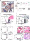
Figure 1. Leptomeninges and brain parenchyma select for distinct metastatic phenotypes
(A) Leptomeningeal metastases (purple cells) enter the CSF via the choroid plexus (upper left) and reside within the CSF where they adhere to the pia mater covering the brain and spinal cord (upper right and lower right). Parenchymal metastases (blue cells) enter the parenchyma through the endothelium, surrounding the vasculature and contacting astrocytes (lower right). (B) Top line: Iterative in vivo selection of leptomeningeal derivative cell lines. 20,000 parental cells are injected into the cisterna magna. After leptomeningeal metastases develop, the mouse is euthanized, cells are collected and grown in culture before injection into a second recipient mouse. This is carried out three times to generate intermediate (Int) cells. 50,000 Int cells are injected intracardially. Mice bearing leptomeningeal metastases are euthanized, cells are collected from the meninges and maintained in culture. These are denoted LeptoM cells. Bottom line: In vivo selection of parenchymal metastatic derivatives. 50,000 parental cells are injected intracardially. Mice with parenchymal metastases are euthanized, brain metastases dissected, dissociated and grown in culture. These cells are injected intracardially once more to generate BrM derivatives. (C–D) 50,000 MDA231 Parental (green), LeptoM (purple) or BrM (blue) cells are injected intracardially into recipient mice. Tumor growth is monitored by BLI once weekly, neuro-anatomic localization (leptomeningeal, c; parenchymal, d) is determined by histology (inset). Scale bar = 100 μm. n = 7 mice Par, 15 mice LeptoM, 10 mice BrM cells. Refer also to Fig. S1. (E) Principle component analysis (PCA) plots of transcriptome from BrM (blue), LeptoM (purple) and Int (orange) cell lines after RNASeq. Genes with base mean ≥ 50 fold change ≥ 2 or ≤ 0.5 and p < 0.01 were included for analysis. (F) Intracranial localization of LeptoM cells after hematogenous dissemination at various time points. 50,000 MDA231-LeptoM cells were injected intracardially on day 0. Three mice were euthanized at each time point. The skull and meninges remained intact throughout tissue processing to maintain architecture. 10 coronal paraffin sections were stained by IHC for GFP; all GFP positive cells were counted on each section as follows choroid plexus (red), leptomeninges (green), parenchyma (blue).
Figure 2
Figure 2. Upregulation of complement component 3 in leptomeningeal metastasis
(A) GSEA analysis of upregulated genes in Par vs. Int and Par vs. LeptoM analyses for each model. KEGG Complement and Coagulation Cascade pathway result is shown. (B) Cancer cell C3 expression by ELISA. Conditioned medium from 1 × 10⁶ cells grown in a 10 cm plate for 48 hours was collected. n = 3 experiments performed in duplicate.
Figure 3
Figure 3. Complement component C3 in human leptomeningeal metastasis
(A) ELISA of CSF obtained from patients with solid tumor primary and clinical symptoms suspicious for leptomeningeal metastasis. Final clinical diagnosis is indicated. n = 37 patients with leptomeningeal metastasis, 20 patients with parenchymal metastases, 12 patients with no CNS metastasis. Refer also to table S1. *** indicates p < 0.001. (B) C3 in CSF by ELISA versus clinical disease burden. Refer also to Figure S3C for calculation of Disease Burden Score. n = 30 patients. ** indicates p < 0.05. (C) C3 in CSF by ELISA versus site of CSF sample. CSF samples were obtained from patients harboring leptomeningeal metastasis, either from lateral ventricle (n = 18) or lumbar cistern (n = 22). *** indicates p < 0.001. (D) C3 expression by qPCR in patient CSF. CD45-depleted cells from CSF were collected from breast and lung cancer patients (n = 5 patients) with leptomeningeal metastasis. Circulating peripheral leukocytes (n = 3), mammary epithelial cells (n = 2) or bronchial epithelial cells (n = 2) were also collected. mRNA is presented as C3:GAPDH on y-axis; B2MG:GAPDH on the × axis. All samples were analyzed in quadruplicate. (E) Immunofluorescence of patient-derived CSF cells for CD45 and C3. n = 6 patients. 100 cells counted per patient. Representative images are presented in (F). **** indicates p < 0.0001. (G) Representative images of primary tumors of patients stained by IHC for cytokeratin and C3. (H) Proportion of total tumor area (as determined by cytokeratin staining) positive for C3 was correlated with clinical outcome. Refer also to figure S3F. n = 76 patients.
Figure 4
Figure 4. C3 is necessary for cancer cell growth within the leptomeninges
(A–B) C3 expression was stably knocked down using two independent short hairpins (shA and shB) or vector control (Ctl) in MDA231-LeptoM derivative cell lines. 2,000 cells were injected intracisternally, BLI was monitored every 5 days. n = 5 mice per group in each of two independent experiments; 10 mice total per group. Histograms represent in vivo BLI imaging at day 28 post inoculation. ***indicates p < 0.001 (C) Kaplan-Meier plot of overall survival of mice injected with MDA231-LeptoM cells with either vCtl or shA as described above. (D) 1,000 MDA231-LeptoM cells were seeded in each well of a tissue-culture treated 96-well plate and allowed to grow in CSF from solid tumor patients with or without LM with 50% artificial CSF. Cell growth was monitored by CellTiter Glo assay at t = 1h and 72h. Data represent two independent experiments performed in quadruplicate. *** indicates p < 0.001 (E–F) 500 MDA231-LeptoM cells were seeded into a 384-well plate containing CSF collected from mice harboring no malignancy. Mice were treated with 1 mg/kg recombinant mouse C3a (rmC3a) or PBS I.P. 30 min prior to CSF collection. For mice treated with PBS, rmC3a was added ex vivo to a final concentration of 20 ng/mL to mouse CSF. Data represent two independent experiments performed in quadruplicate. *** indicates p < 0.001 (G) 2,000 Parental cells were introduced into the cisterna magna with either recombinant C3a or vehicle. Additional rmC3a or vehicle was delivered intracisternally every 7 days. (H) BLI at day 14 of mice inoculated with MDA231 Parental cells, treated as described in (G). * indicates p < 0.01 (I) Kaplan-Meier plot of overall survival of mice in (H). n = 10 mice per group.
Figure 5
Figure 5. C3 perturbs choroid plexus barrier function
(A) Immunofluorescence of C57/Bl6 mouse choroid plexus for C3aR (red) or SPAK (green), a marker of the luminal CPEpi surface. Scale bar represents 100 μm. (B) Human CPEpi cells are grown on poly-L-Lysine coated transwells until TEER is greater than 200 Ω/cm2. Media was changed to conditioned media (CM) as indicated and TEER measurements were obtained every 12 hours. Two independent experiments performed in triplicate are averaged. (C) Conditioned media from MDA231 parental cells supplemented with recombinant mouse C3a at 5 ng/mL (rmC3a), open circles or equal volume PBS, closed circles. (D) Conditioned media from MDA231-LeptoM derivative cells immunodepleted with anti-C3 (open circles), vehicle (closed circles) or isotype control (grey circles). (E–G) C57/Bl6 mice (C3aR+/+ or C3aR−/) without malignancy were treated with 1 mg/mL rmC3a I.P. or vehicle alone prior to intracardiac introduction of mixed dextrans (Cascade Blue-10 kDa, FITC-40 kDa, Texas Red-500 kDa 1:1:1 in PBS). CSF and peripheral blood sampling occurred 30 min later. n = 5 mice, each fluorescence measurement performed in triplicate. *** p < 0.001; **** p < 0.0001; NS = not significant (H–I) Choroid plexus from the lateral ventricles of C57/Bl6 mice were treated 37°C with conditioned media from LLC Parental cells supplemented with 10 ng/mL rmC3a or vehicle alone. After 0, 30 or 120 minutes at 37C, specimens were lysed for western blotting (H). After three hours at 37C, specimens were either fixed for IF (G) against ZO-1 (red) and claudin (green) nuclei are marked with DAPI (blue). (J–K) Migration of 20,000 serum-starved Par cells, LeptoM control short hairpin (vCtl) or C3 knock down short hairpin (shA) across a confluent monolayer of human CPEpi cells toward artificial CSF + 1% FBS. Cells were allowed to migrate 12 hours before quantification of cells. Two independent experiments were performed in duplicate. Ten high-power fields (40×) were counted per condition per replicate. NS = not significant.
Figure 6
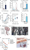
Figure 6. C3a mediates amphiregulin influx to adapt CSF for cancer cell growth
(A) 2,000 LLC-LeptoM cells were introduced intracisternally into C3aR−/− or C3aR +/+ mice in C57/Bl6 background. n = 5 −/− mice, 10 +/+ mice. * p < 0.05 (B) ELISA for human amphiregulin of CSF obtained from patients with solid tumor primary and clinical symptoms suspicious for leptomeningeal metastasis. Final clinical diagnosis is indicated. n = 11 patients with leptomeningeal metastasis, 11 patients with no CNS metastasis. *** p < 0.001 (C) 1,000 MDA-LeptoM cells were seeded in each well of a tissue-culture treated 96-well plate and allowed to grow in artificial CSF supplemented with recombinant amphiregulin. Cell growth was monitored by CellTiter-Glo assay at 72 h. Data represent two independent experiments performed in quadruplicate. *** p < 0.001 (D) 1,000 PC9-LeptoM cells were seeded in each well of a tissue-culture treated 96-well plate and allowed to grow in artificial CSF supplemented with recombinant amphiregulin. Cell growth was monitored by CellTiter-Glo assay at the indicated time points. Data represent two independent experiments performed in quadruplicate. (E) 2,000 MDA Par cells were introduced into the cisterna magna with either recombinant amphiregulin or vehicle. Additional amphiregulin or vehicle was delivered intracisternally every 7 days. BLI on day 14 is illustrated. * p < 0.05; *** p < 0.001 (F) Naïve C57/Bl6 mice were treated with either 10 ng/mL rmC3a or vehicle prior to CSF and blood sampling. n = 3 mice in each treatment group. Amphiregulin was assayed by ELISA in serum and CSF from each mouse in triplicate. Data are presented as ratio of CSF Amphiregulin : Serum Amphiregulin; * p < 0.05 (G) Patients with leptomeningeal metastasis and intention to treat with intra-ventricular trastuzumab have an intra-ventricular (Ommaya) reservoir placed in the lateral ventricles. CSF adjacent to the choroid plexus is sampled just prior to treatment and analyzed for C2, C3, C4, Factor D and amphiregulin (AR) by ELISA. T1 post-contrast MRI from Patient 1 demonstrating clinical response is shown at right. Deposits of leptomeningeal metastasis appear as white plaques over the cervical spine and are indicated by red arrowheads. (H) Analysis of CSF from Patient 1 for indicated analytes at three time points. (I) Cancer cells within the cerebrospinal fluid produce C3. C3a binds to and activates C3aR on the choroid plexus leading to loosening of tight junctions and impaired barrier function. Select plasma components, including amphiregulin, gain entry to the cerebrospinal fluid where they support cancer cell growth.
Figure 7
Figure 7. C3aR as a therapeutic target in leptomeningeal metastasis
(A) 2,000 MDA231-LeptoM cells were introduced into the cisterna magna on day 0. Mice were treated with 10 mg/kg C3aR agonist (Ag), 10 mg/kg antagonist (Ant) or vehicle (Veh) I.P. twice weekly, tumor cell growth was monitored by BLI. n = 10 mice per group. **** p < 0.0001 (B) Survival analysis of mice treated in (A). (C) 2,000 MDA231-LeptoM, PC9-LeptoM, HCC1954-LeptoM or LCC-LeptoM cells were introduced into the CSF on day 0 and treated with Veh or Ant as described in (A). n = 10 mice per group. BLI on day 14 is illustrated. * p < 0.05; ** p < 0.01; *** p < 0.001.

Comment in

  • Metastasis: Breaching barriers.
    Dart A. Dart A. Nat Rev Cancer. 2017 May;17(5):270. doi: 10.1038/nrc.2017.27. Epub 2017 Apr 7. Nat Rev Cancer. 2017. PMID: 28386090 No abstract available.

References

    1. Acharyya S, Oskarsson T, Vanharanta S, Malladi S, Kim J, Morris PG, Manova-Todorova K, Leversha M, Hogg N, Seshan VE, et al. A CXCL1 paracrine network links cancer chemoresistance and metastasis. Cell. 2012;150:165–178. - PMC - PubMed
    1. Addison CL, Ding K, Zhao H, Le Maitre A, Goss GD, Seymour L, Tsao MS, Shepherd FA, Bradbury PA. Plasma transforming growth factor alpha and amphiregulin protein levels in NCIC Clinical Trials Group BR.21. J Clin Oncol. 2010;28:5247–5256. - PubMed
    1. Ames RS, Lee D, Foley JJ, Jurewicz AJ, Tornetta MA, Bautsch W, Settmacher B, Klos A, Erhard KF, Cousins RD, et al. Identification of a selective nonpeptide antagonist of the anaphylatoxin C3a receptor that demonstrates antiinflammatory activity in animal models. J Immunol. 2001;166:6341–6348. - PubMed
    1. Ames RS, Li Y, Sarau HM, Nuthulaganti P, Foley JJ, Ellis C, Zeng Z, Su K, Jurewicz AJ, Hertzberg RP, et al. Molecular cloning and characterization of the human anaphylatoxin C3a receptor. J Biol Chem. 1996;271:20231–20234. - PubMed
    1. An YJ, Cho HR, Kim TM, Keam B, Kim JW, Wen H, Park CK, Lee SH, Im SA, Kim JE, et al. An NMR metabolomics approach for the diagnosis of leptomeningeal carcinomatosis in lung adenocarcinoma cancer patients. Int J Cancer. 2015;136:162–171. - PubMed

Publication types

Substances