

Developmental, Genetic, Dietary, and Xenobiotic Influences on Neonatal Hyperbilirubinemia
- PMID: 28283555
- PMCID: PMC5416747
- DOI: 10.1124/mol.116.107524
Developmental, Genetic, Dietary, and Xenobiotic Influences on Neonatal Hyperbilirubinemia
Abstract
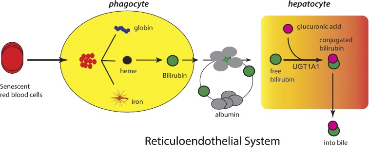
Hyperbilirubinemia, caused by the accumulation of unconjugated bilirubin, is one of the most common clinical diagnoses in both premature and term newborns. Owing to the fact that bilirubin is metabolized solely through glucuronidation by UDP-glucuronosyltransferase (UGT) 1A1, it is now known that immaturity of UGT1A1, in combination with the overproduction of bilirubin during the developmental stage, acts as a bottleneck to bilirubin elimination and predisposes the infant to high total serum bilirubin levels. Although neonatal jaundice is mostly benign, excessively high levels of serum bilirubin in a small percentage of newborns can cause bilirubin-induced neurologic dysfunction, potentially leading to permanent brain damage, a condition known as kernicterus Although a large portion of hyperbilirubinemia cases in newborns are associated with hemolytic diseases, we emphasize here the impaired ability of UGT1A1 to eliminate bilirubin that contributes to hyperbilirubinemia-induced neurotoxicity in the developmental stage. As a series of hereditary UGT1A1 mutations have been identified that are associated with UGT1A1 deficiency, new evidence has verified that delayed expression of UGT1A1 during the early stages of neonatal development is a tightly controlled event involving coordinated intrahepatic and extrahepatic regulation. This review recapitulates the progress that has been made in recent years in understanding the causes and physiopathology of severe hyperbilirubinemia, investigating molecular mechanisms underlying bilirubin-induced encephalopathy, and searching for potential therapies for treating pathologic hyperbilirubinemia. Several animal models have been developed to make it possible to examine bilirubin-induced neurotoxicity from multiple directions. Moreover, environmental factors that may alleviate or worsen the condition of hyperbilirubinemia are discussed.
Copyright © 2017 by The American Society for Pharmacology and Experimental Therapeutics.
Figures

Similar articles
-
Developmental hyperbilirubinemia and CNS toxicity in mice humanized with the UDP glucuronosyltransferase 1 (UGT1) locus.Proc Natl Acad Sci U S A. 2010 Mar 16;107(11):5024-9. doi: 10.1073/pnas.0913290107. Epub 2010 Mar 1. Proc Natl Acad Sci U S A. 2010. PMID: 20194756 Free PMC article.
-
Glucose induces intestinal human UDP-glucuronosyltransferase (UGT) 1A1 to prevent neonatal hyperbilirubinemia.Sci Rep. 2014 Sep 11;4:6343. doi: 10.1038/srep06343. Sci Rep. 2014. PMID: 25209391 Free PMC article.
-
Reduced expression of UGT1A1 in intestines of humanized UGT1 mice via inactivation of NF-κB leads to hyperbilirubinemia.Gastroenterology. 2012 Jan;142(1):109-118.e2. doi: 10.1053/j.gastro.2011.09.045. Epub 2011 Oct 6. Gastroenterology. 2012. PMID: 21983082 Free PMC article.
-
Role of extrahepatic UDP-glucuronosyltransferase 1A1: Advances in understanding breast milk-induced neonatal hyperbilirubinemia.Toxicol Appl Pharmacol. 2015 Nov 15;289(1):124-32. doi: 10.1016/j.taap.2015.08.018. Epub 2015 Sep 2. Toxicol Appl Pharmacol. 2015. PMID: 26342858 Free PMC article. Review.
-
Molecular diagnostic update in hereditary hemolytic anemia and neonatal hyperbilirubinemia.Int J Lab Hematol. 2019 May;41 Suppl 1:95-101. doi: 10.1111/ijlh.13014. Int J Lab Hematol. 2019. PMID: 31069991 Review.
Cited by
-
Developing and evaluating a predictive model for neonatal hyperbilirubinemia based on UGT1A1 gene polymorphism and clinical risk factors.Front Pediatr. 2024 Feb 29;12:1345602. doi: 10.3389/fped.2024.1345602. eCollection 2024. Front Pediatr. 2024. PMID: 38487473 Free PMC article.
-
Impact of Low Birth Weight and Prematurity on Neonatal Raltegravir Pharmacokinetics: Impaact P1097.J Acquir Immune Defic Syndr. 2020 Dec 15;85(5):626-634. doi: 10.1097/QAI.0000000000002492. J Acquir Immune Defic Syndr. 2020. PMID: 32925360 Free PMC article.
-
Identification of Genetic Risk Factors for Neonatal Hyperbilirubinemia in Fujian Province, Southeastern China: A Case-Control Study.Biomed Res Int. 2018 Sep 12;2018:7803175. doi: 10.1155/2018/7803175. eCollection 2018. Biomed Res Int. 2018. PMID: 30298137 Free PMC article.
-
Humanized UGT1 Mice, Regulation of UGT1A1, and the Role of the Intestinal Tract in Neonatal Hyperbilirubinemia and Breast Milk-Induced Jaundice.Drug Metab Dispos. 2018 Nov;46(11):1745-1755. doi: 10.1124/dmd.118.083212. Epub 2018 Aug 9. Drug Metab Dispos. 2018. PMID: 30093417 Free PMC article.
-
Genetic alterations and molecular mechanisms underlying hereditary intrahepatic cholestasis.Front Pharmacol. 2023 May 31;14:1173542. doi: 10.3389/fphar.2023.1173542. eCollection 2023. Front Pharmacol. 2023. PMID: 37324459 Free PMC article. Review.
References
-
- Barateiro A, Miron VE, Santos SD, Relvas JB, Fernandes A, Ffrench-Constant C, Brites D. (2013) Unconjugated bilirubin restricts oligodendrocyte differentiation and axonal myelination. Mol Neurobiol 47:632–644. - PubMed
-
- Bellodi-Privato M, Aubert D, Pichard V, Myara A, Trivin F, Ferry N. (2005) Successful gene therapy of the Gunn rat by in vivo neonatal hepatic gene transfer using murine oncoretroviral vectors. Hepatology 42:431–438. - PubMed
-
- Bhutani VK, Johnson L. (2009) Kernicterus in the 21st century: frequently asked questions. J Perinatol 29 (Suppl 1):S20–S24. - PubMed
Publication types
MeSH terms
Substances
Grants and funding
LinkOut - more resources
Full Text Sources
Other Literature Sources
Research Materials
