. 2017 Apr 12;37(15):4128-4144.
doi: 10.1523/JNEUROSCI.3193-16.2017. Epub 2017 Mar 10.

Gene Expression Profiling with Cre-Conditional Pseudorabies Virus Reveals a Subset of Midbrain Neurons That Participate in Reward Circuitry

Affiliations

Gene Expression Profiling with Cre-Conditional Pseudorabies Virus Reveals a Subset of Midbrain Neurons That Participate in Reward Circuitry

Lisa E Pomeranz et al. J Neurosci. .

Abstract

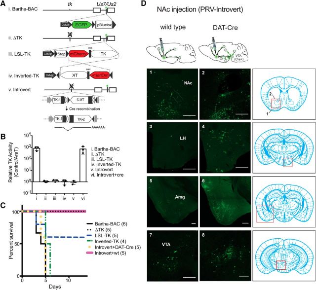
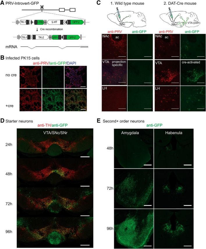
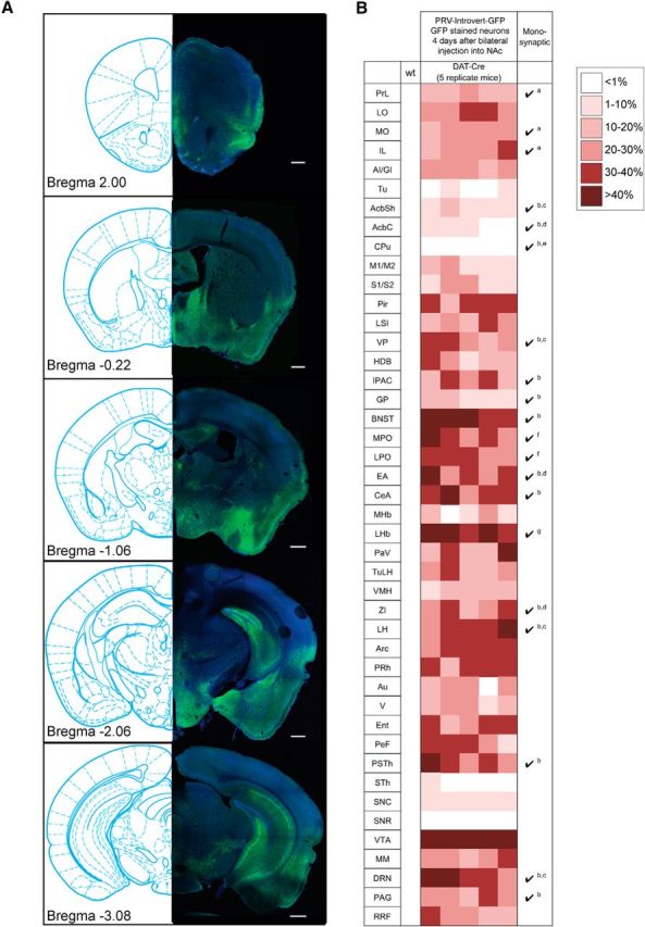
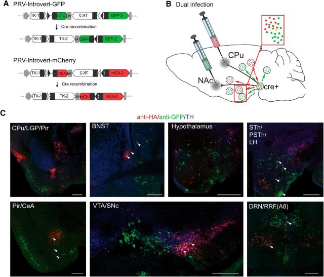
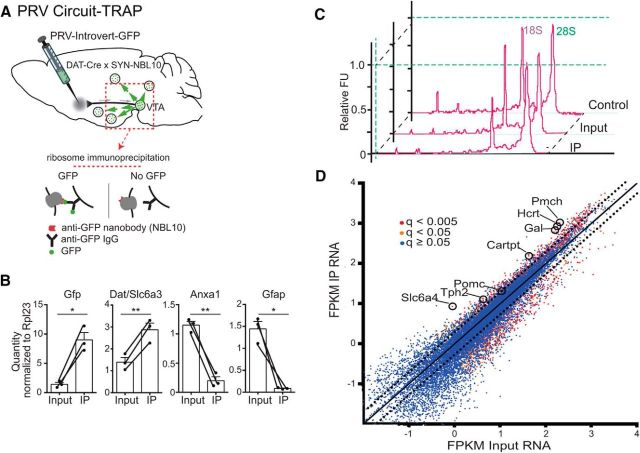
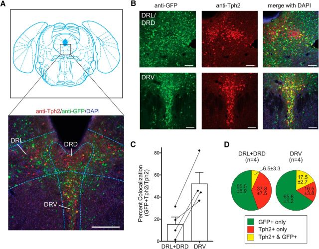
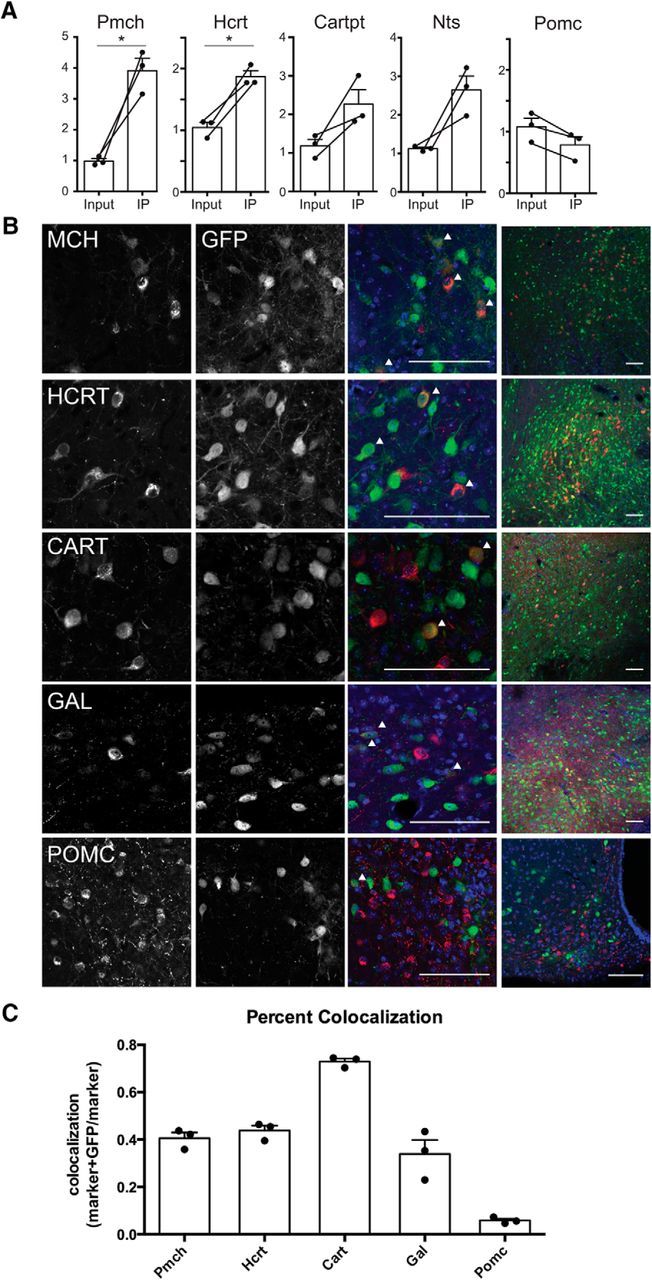
The mesolimbic dopamine pathway receives inputs from numerous regions of the brain as part of a neural system that detects rewarding stimuli and coordinates a behavioral response. The capacity to simultaneously map and molecularly define the components of this complex multisynaptic circuit would thus advance our understanding of the determinants of motivated behavior. To accomplish this, we have constructed pseudorabies virus (PRV) strains in which viral propagation and fluorophore expression are activated only after exposure to Cre recombinase. Once activated in Cre-expressing neurons, the virus serially labels chains of presynaptic neurons. Dual injection of GFP and mCherry tracing viruses simultaneously illuminates nigrostriatal and mesolimbic circuitry and shows no overlap, demonstrating that PRV transmission is confined to synaptically connected neurons. To molecularly profile mesolimbic dopamine neurons and their presynaptic inputs, we injected Cre-conditional GFP virus into the NAc of (anti-GFP) nanobody-L10 transgenic mice and immunoprecipitated translating ribosomes from neurons infected after retrograde tracing. Analysis of purified RNA revealed an enrichment of transcripts expressed in neurons of the dorsal raphe nuclei and lateral hypothalamus that project to the mesolimbic dopamine circuit. These studies identify important inputs to the mesolimbic dopamine pathway and further show that PRV circuit-directed translating ribosome affinity purification can be broadly applied to identify molecularly defined neurons comprising complex, multisynaptic circuits.SIGNIFICANCE STATEMENT The mesolimbic dopamine circuit integrates signals from key brain regions to detect and respond to rewarding stimuli. To further define this complex multisynaptic circuit, we constructed a panel of Cre recombinase-activated pseudorabies viruses (PRVs) that enabled retrograde tracing of neural inputs that terminate on Cre-expressing neurons. Using these viruses and Retro-TRAP (translating ribosome affinity purification), a previously reported molecular profiling method, we developed a novel technique that provides anatomic as well as molecular information about the neural components of polysynaptic circuits. We refer to this new method as PRV-Circuit-TRAP (PRV circuit-directed TRAP). Using it, we have identified major projections to the mesolimbic dopamine circuit from the lateral hypothalamus and dorsal raphe nucleus and defined a discrete subset of transcripts expressed in these projecting neurons, which will allow further characterization of this important pathway. Moreover, the method we report is general and can be applied to the study of other neural circuits.

Keywords: RNASeq; mesolimbic; pseudorabies virus.


Figures

Figure 1.
Figure 1.
Cre-conditional retrograde tracing with PRV-Introvert. A, Schematic of PRV-Introvert construction. Bartha-BAC, the parent of PRV-Introvert, expresses full length PRV tk from its native promoter (i). EGFP (green asterisk) and the sequence for maintenance as a bacterial artificial chromosome (pBluelox; Smith and Enquist, 2000) are inserted into the Us7/Us2 locus. ΔTK has a deletion of the PRV tk gene (ii). In LSL-TK, PRV tk is controlled by a lox-stop-lox cassette between the CMV immediate early (IE) promoter and mCherry-ires-tk, all of which is inserted into the PRV gG/US4 locus (iii). In Inverted-tk, mCherry-ires-tk is reversed between two lox sites (iv). In PRV-Introvert, the PRV tk gene is split into 5′ and 3′ ends between two sets of inverted lox sites. Cre recombination flips the 3′ end of PRV tk into the same orientation as the 5′ end and removes two lox sites. The single lox site that remains within the PRV tk transcript is removed by splicing. Open triangles represent loxN sites while lox2722 sites are represented by shaded triangles (v). B, TK activity of parental strain (Bartha-BAC) and mutants. Virus stocks were titered in the presence or absence of AraT, a nucleoside analog that inhibits viral replication in the presence of PRV TK. TK activity is measured as the ratio of the number of plaques without AraT/ with AraT. C, Kaplan–Meier curve showing mouse survival after injection of PRV into the NAc. Bartha-BAC, ΔTK, LSL-TK, Inverted-TK, and Introvert + wt show data for wild-type mice. Introvert + DAT-Cre shows survival of DAT-Cre mice after NAc injection of PRV-Introvert-GFP. Numbers in parentheses are the number of mice injected with each virus. D, PRV-Introvert injected into the NAc of wild-type (1, 3, 5, 7) or DAT-Cre (2, 4, 6, 8). Four days after injection, the brains were perfused, sectioned, and stained with anti-GFP. Scale bars: 250 μm.
Figure 2.
Figure 2.
PRV-Introvert-GFP does not express GFP or spread in the CNS without Cre recombinase. A, Cre-dependent expression of PRV tk and GFP using a FLEX intron and 2A ribosomal skip sequence. Exposure to Cre inverts the 3′ exon of PRV tk (TK-2) and the 5′ exon of GFP (GFP-1) and excises one loxN (open triangles) and one lox2722 site (shaded triangles). The remaining lox sites are removed by splicing. B, Cre recombinase is required for GFP expression during PRV-Introvert-GFP infection of PK15 cells. C, PRV-Introvert-GFP injection of wild-type and DAT-Cre mice. 1, Wild-type mouse showing limited expression of viral proteins and no GFP three days after NAc injection with PRV-Introvert-GFP. 2, Representative sections from DAT-Cre mouse four days post NAc injection. ac: anterior commissure. D, Time course of PRV spread in DAT-Cre 24, 48, 72, and 96 h after bilateral injection in the NAc. Anti-TH staining marks dopamine neurons. E, Structures infected 48, 72, and 96 h after infection that represent second or higher order connections to DAT-Cre starter cells in the VTA. Scale bars: B, 100 μm; C, 250 μm; D, E, 500 μm.
Figure 3.
Figure 3.
Main afferent targets of mesolimbic dopamine circuitry. A, Representative anti-GFP staining of structures that exhibit GFP reactivity after Cre-dependent retrograde tracing. Scale bars: 500 μm. B, Summary of tracing data 4 d after infection. Shown are one wild-type and five DAT-cre mice injected in the NAc PRV-Introvert-GFP. No anti-GFP reactivity was detected in a wild-type mouse injected as a control. Monosynaptic connectivity of structures was established in previous studies as indicated: a, Monosynaptic connection from anterior cortex to VTA dopamine neurons projecting to the lateral NAc (Beier et al., 2015); b, number of input neurons ≥200/10,000 total inputs to VTA dopamine neurons (Watabe-Uchida et al., 2012); c, monosynaptic connections to both medial and lateral NAc-projecting VTA dopamine neurons ≥5% (Beier et al., 2015); d, monosynaptic connections to lateral NAc-projecting VTA dopamine neurons ≥5% (Beier et al., 2015); e, monosynaptic connections from dorsal striatum to lateral NAc-projecting VTA dopamine neurons (Beier et al., 2015); f, monosynaptic connections from the preoptic area to both medial and lateral NAc-projecting VTA dopamine neurons (Beier et al., 2015); g, monosynaptic connections to medial NAc-projecting VTA dopamine neurons ≥5% (Beier et al., 2015). PrL, Prelimbic cortex; LO, lateral orbital cortex; MO, medial orbital cortex; IL, infralimbic cortex; AI/GI, insular cortex (granular/agranular); Tu, olfactory tubercle; AcbSh, nucleus accumbens shell; AcbC, nucleus accumbens core; M1/M2, motor cortex (primary/secondary); S1/S2, somatosensory cortex (primary/secondary); Pir, piriform cortex; LSI, lateral intermediate septal nucleus; VP, ventral pallidum; HDB, nucleus of the horizontal limb of the diagonal band; IPAC, interstitial nucleus of the posterior limb of the anterior commissure; GP, globus pallidus; MPO, medial preoptic nucleus; LPO, lateral preoptic nucleus; EA, sublenticular extended amygdala; CeA central amygdaloid nucleus; MHb, medial habenular nucleus; PaV, paraventricular hypothalamic nucleus; TuLH, tuberal region of the hypothalamus; VMH, ventromedial hypothalamic nucleus; ZI, zona incerta; PRh, perirhinal cortex; Au, auditory cortex; V, visual cortex; Ent, entorhinal cortex; PeF Perifornical nucleus; PSTh, parasubthalamic nucleus; STh, subthalamic nucleus; SNC, substantia nigra compact part; SNR, substantia nigra reticular part; MM, supramammilary nucleus; PAG, periaqueductal gray; RRF, retrorubral field.
Figure 4.
Figure 4.
Dual tracing of inputs to CPu- and NAc-projecting dopamine neurons shows specificity of retrograde labeling and minimal cross talk between circuitry. A, PRV-Introvert-mCherry is identical to PRV-Introvert-GFP except that GFP has been replaced with HA-mCherry. B, Dual injection in the NAc and CPu with isogenic strains of PRV-Introvert results in only a few neurons infected with both viruses (yellow). C, PRV-Introvert-GFP and PRV-Introvert-mCherry were injected into the right NAc and CPu, respectively, of DAT-Cre mice. Four days after injection, brains were assayed for anti-HA (red) and anti-GFP (green) staining. Arrowheads denote neurons expressing both red and green label. Scale bars: 500 μm. Abbreviations are defined in the Figure 3 legend.
Figure 5.
Figure 5.
PRV-Circuit-TRAP of hypothalamic and midbrain neurons projecting to the mesolimbic dopamine circuit. A, Strategy for PRV-Circuit-TRAP. PRV-Introvert-GFP was injected bilaterally into the NAc of DAT-Cre × SYN-NBL10 mice. Translating ribosomes tagged with GFP binding nanobody (NBL10) were immunoprecipitated from a section of midbrain 3 days after injection of PRV-Introvert-GFP. B, Results of qPCR of input and IP samples from PRV-Circuit-TRAP for Gfp (p < 0.05), Dat/Slc6a3 (p < 0.01), Anxa1 (p < 0.01), and Gfap (p < 0.05). Fold enrichment for each gene from the IP or input sample is normalized to the housekeeping gene Rpl23. C, Electropherogram of representative control, input, and IP RNA samples measured on an Agilent 2100 Bioanalyzer before sequencing on an Illumina HiSeq 2000. D, Differential enrichment of immunoprecipitated transcripts. Dotted lines indicate twofold enrichment or depletion in IP RNA compared with input RNA. Colors of dots indicate the significance of enrichment: red, q < 0.005; orange, q < 0.05; blue, q ≥ 0.05. *p < 0.05; **p < 0.01.
Figure 6.
Figure 6.
Serotonergic effectors of mesolimbic dopamine neurons. A, Coronal section showing Bregma −4.72 (Franklin and Paxinos, 2008) and section of infected CNS tissue indicating the dorsomedial, ventromedial, and dorsolateral wings of the dorsal raphe. The section is stained with anti-Tph2, anti-GFP, and DAPI. B, High-magnification images of DRL/DRD and DRV. C, Percent colocalization (ratio of the number of cells in the DRN stained with both Tph2 and GFP to the number stained with Tph2 only). Error bars indicate SEM. D, Quantification of cells in the DRN stained with Tph2 only, GFP only, or both Tph2 and GFP. Scale bars: A, 250 μm; B, 100 μm.
Figure 7.
Figure 7.
Hypothalamic effectors of mesolimbic dopamine neurons express appetitive markers. A, qPCR of IP and input samples from PRV-Circuit-TRAP. Pmch (p = 0.024) and Hcrt (p = 0.0134) RNA were significantly increased in IP compared with input samples. Cart (p = 0.0981), Nts (p = 0.0627), and Pomc (p = 0.054) IP and input RNA are not significantly different. B, Immunohistochemistry of neuronal markers and GFP in PRV-Introvert-GFP-infected CNS. Arrowheads indicate cells stained with both marker and GFP. Scale bars, 100 μm. C, Quantification of PRV-Introvert-GFP colocalization with appetitive markers shown as number of neurons coexpressing marker and GFP out of the total number of marker-expressing neurons. Percentages are shown as ±SEM. *p < 0.05.

Similar articles

Cited by

References

    1. Anthony TE, Dee N, Bernard A, Lerchner W, Heintz N, Anderson DJ (2014) Control of stress-induced persistent anxiety by an extra-amygdala septohypothalamic circuit. Cell 156:522–536. 10.1016/j.cell.2013.12.040 - DOI - PMC - PubMed
    1. Ashburner M, Ball CA, Blake JA, Botstein D, Butler H, Cherry JM, Davis AP, Dolinski K, Dwight SS, Eppig JT, Harris MA, Hill DP, Issel-Tarver L, Kasarskis A, Lewis S, Matese JC, Richardson JE, Ringwald M, Rubin GM, Sherlock G (2000) Gene ontology: tool for the unification of biology. The Gene Ontology Consortium. Nat Genet 25:25–29. 10.1038/75556 - DOI - PMC - PubMed
    1. Banfield BW, Kaufman JD, Randall JA, Pickard GE (2003) Development of pseudorabies virus strains expressing red fluorescent proteins: new tools for multisynaptic labeling applications. J Virol 77:10106–10112. 10.1128/JVI.77.18.10106-10112.2003 - DOI - PMC - PubMed
    1. Beier KT, Saunders A, Oldenburg IA, Miyamichi K, Akhtar N, Luo L, Whelan SP, Sabatini B, Cepko CL (2011) Anterograde or retrograde transsynaptic labeling of CNS neurons with vesicular stomatitis virus vectors. Proc Natl Acad Sci U S A 108:15414–15419. 10.1073/pnas.1110854108 - DOI - PMC - PubMed
    1. Beier KT, Borghuis BG, El-Danaf RN, Huberman AD, Demb JB, Cepko CL (2013) Transsynaptic tracing with vesicular stomatitis virus reveals novel retinal circuitry. J Neurosci 33:35–51. 10.1523/JNEUROSCI.0245-12.2013 - DOI - PMC - PubMed

Publication types