. 2017 Mar 9;9(3):823-851.
doi: 10.18632/aging.101195.

Amyotrophic lateral sclerosis, gene deregulation in the anterior horn of the spinal cord and frontal cortex area 8: implications in frontotemporal lobar degeneration

Affiliations

Amyotrophic lateral sclerosis, gene deregulation in the anterior horn of the spinal cord and frontal cortex area 8: implications in frontotemporal lobar degeneration

Pol Andrés-Benito et al. Aging (Albany NY). .

Abstract

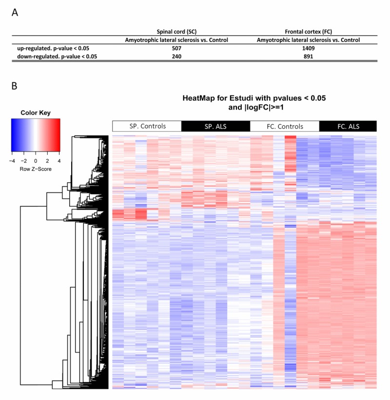
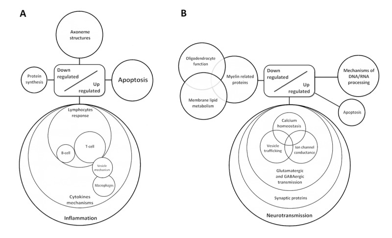
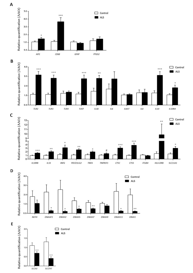
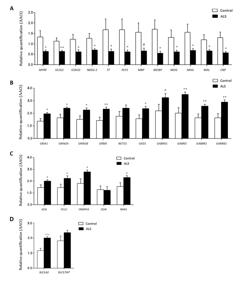
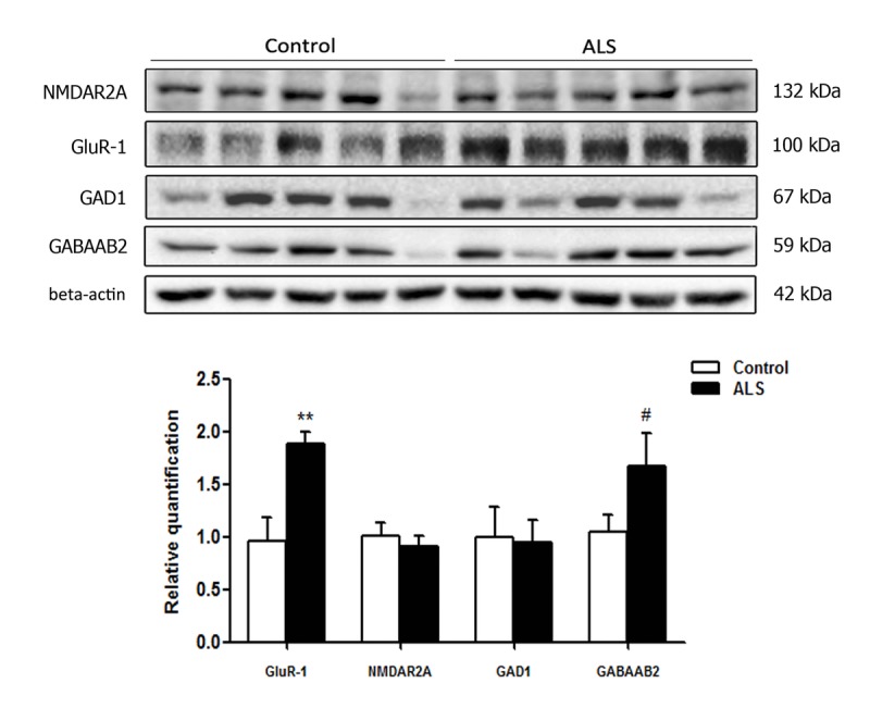
Transcriptome arrays identifies 747 genes differentially expressed in the anterior horn of the spinal cord and 2,300 genes differentially expressed in frontal cortex area 8 in a single group of typical sALS cases without frontotemporal dementia compared with age-matched controls. Main up-regulated clusters in the anterior horn are related to inflammation and apoptosis; down-regulated clusters are linked to axoneme structures and protein synthesis. In contrast, up-regulated gene clusters in frontal cortex area 8 involve neurotransmission, synaptic proteins and vesicle trafficking, whereas main down-regulated genes cluster into oligodendrocyte function and myelin-related proteins. RT-qPCR validates the expression of 58 of 66 assessed genes from different clusters. The present results: a. reveal regional differences in de-regulated gene expression between the anterior horn of the spinal cord and frontal cortex area 8 in the same individuals suffering from sALS; b. validate and extend our knowledge about the complexity of the inflammatory response in the anterior horn of the spinal cord; and c. identify for the first time extensive gene up-regulation of neurotransmission and synaptic-related genes, together with significant down-regulation of oligodendrocyte- and myelin-related genes, as important contributors to the pathogenesis of frontal cortex alterations in the sALS/frontotemporal lobar degeneration spectrum complex at stages with no apparent cognitive impairment.

Keywords: amyotrophic lateral sclerosis; excitotoxicity; frontal cortex; frontotemporal lobar degeneration; neuroinflammation; spinal cord.


Conflict of interest statement

CONFLICTS OF INTEREST

The authors declare no conflict of interest.

Figures

Figure 1
Figure 1
(A) Total number of significantly different expressed genes comparing transcriptomic profiles between groups and regions. (B) Hierarchical clustering heat map of expression intensities of mRNA array transcripts reflect differential gene expression profiles in the anterior horn of the spinal cord and frontal cortex area 8 in ALS compared with controls. Differences between groups are considered statistically significant at p-value ≤ 0.05. Abbreviations: ALS: amyotrophic lateral sclerosis; FC: frontal cortex area 8; mRNA: messenger RNA; SP: anterior horn of the spinal cord lumbar level
Figure 2
Figure 2
Diagram showing de-regulated gene clusters in the anterior horn of the spinal cord (A) and frontal cortex area 8 in ALS (B) as revealed by whole transcriptome arrays.
Figure 3
Figure 3
mRNA expression levels of selected deregulated genes identified by microarray analysis in the anterior horn of the spinal cord in ALS determined by TaqMan RT-qPCR assays. (A) general glial markers; (B-C) mediators of the inflammatory response; and (D) axolemal components. Up of AIGF1 and CD68, toll-like receptors, cytokines and receptors, chemokines and other mediators of the innate and adaptative inflammatory responses. Axolemal genes, excepting NEFH, which shows a non-significant trend to decrease, are significantly down-regulated. (E) glutamate transporter coding genes. The significance level is set at * p < 0.05, ** p < 0.01 and *** p < 0.001.
Figure 4
Figure 4
mRNA expression levels of selected deregulated genes identified by microarray analysis in frontal cortex area 8 of ALS cases determined by TaqMan RT-qPCR assays. (A) oligodendroglial and myelin-related genes; (B) glutamatergic and GABAergic-related genes and corresponding ionotropic and metabotropic receptors; (C) genes coding for synaptic cleft proteins. Significant up of genes linked to neurotransmission and synapses, and significant down of genes linked to oligodendroglia and myelination. (D) Glutamate transporter coding genes. The significance level is set at * p < 0.05, ** p < 0.01 and *** p < 0.001, and tendencies at # < 0.1.
Figure 5
Figure 5
Anterior horn of the spinal cord. Haematoxilin and eosin staining showing damaged neurons in ALS (a). Immuno-histochemistry to TDP-43 showing skein-like intracytoplasmic inclusions (b), VDAC (c, d), GFAP (e, f), IBA-1 (g, h), CD68 (i, j), HLA-DRB1 (k, l), HLA-DRB5 (m, n), IL-10 (o, p), TNF-α (q, r) and GluT (SLC1A2) (s, t) in the anterior horn of the lumbar spinal cord in control (c, e, g, I, k, m, o, q, s) and sALS (a, b, d, f, h, j, l, n, p, r, t) cases. TDP-43-immunoreactive cytoplasmic inclusions are seen in motor neurons in sALS. GFAP is increased in reactive astrocytes; microglial cells have a round, amoeboid morphology as seen with IBA-1, CD-68, HLA-DRB1, and HLA-DRB5 antibodies. VDAC immunoreactivity is decreased whereas IL-10 and TNF-α is increased in remaining motor neurons in sALS. SLC1A2 immunoreactivity is reduced in the membrane of neurons and in neuropil of the anterior horn in sALS. Paraffin sections, slightly counterstained with haematoxylin; a, c-d, o-t, bar in t = 40μm; e-n, bar in = 20μm; bar in b = 10μm
Figure 6
Figure 6
Gel electrophoresis and western blotting to glutamate receptor ionotropic, NMDA 2A (NMDAR2A), α‐amino‐3‐hydroxy‐5‐methyl‐4‐isoxazolepropionic acid receptor 1 (GluR‐1), glutamate decarboxylase 1 (GAD1) and gamma‐aminobutyric acid receptor subunit beta‐2 (GABAAB2) in the frontal cortex area 8 of control and ALS. Significant increased levels of GluR‐1 and a tendency to increased levels of GABAAB2 are seen in ALS when compared with controls. The significance level is set at ** p < 0.01 and tendencies at # < 0.1.

References

    1. Horobágyi T, Cairns NJ. Amyotrophic lateral sclerosis and frontotemporal lobar degeneration. In: Kovacs GG, editor. Neuropathology of neurodegenerative diseases: a practical guide. Cambridge Press; Cambridge: 2015. pp. 209–248.
    1. Strong MJ, Hortobágyi T, Okamoto K, Kato S. Amyotrophic lateral sclerosis, primary lateral sclerosis, and spinal muscular atrophy. In: Dickson DW, editor. Neurodegeneration: the molecular pathology of dementia and movement disorders. 2nd. Wiley-Blackwell; Oxford: 2011. pp. 418–433.
    1. Boillée S, Vande Velde C, Cleveland DW. ALS: a disease of motor neurons and their nonneuronal neighbors. Neuron. 2006;52:39–59. doi: 10.1016/j.neuron.2006.09.018. - DOI - PubMed
    1. Calvo A, Moglia C, Balma M, Chiò A. Involvement of immune response in the pathogenesis of amyotrophic lateral sclerosis: a therapeutic opportunity? CNS Neurol Disord Drug Targets. 2010;9:325–30. doi: 10.2174/187152710791292657. - DOI - PubMed
    1. D'Amico E, Factor-Litvak P, Santella RM, Mitsumoto H. Clinical perspective on oxidative stress in sporadic amyotrophic lateral sclerosis. Free Radic Biol Med. 2013;65:509–27. doi: 10.1016/j.freeradbiomed.2013.06.029. - DOI - PMC - PubMed

Publication types