

Amygdala enlargement: Temporal lobe epilepsy subtype or nonspecific finding?
- PMID: 28284051
- PMCID: PMC5945291
- DOI: 10.1016/j.eplepsyres.2017.02.019
Amygdala enlargement: Temporal lobe epilepsy subtype or nonspecific finding?
Abstract
Objective: Amygdala enlargement (AE) is observed in patients with temporal lobe epilepsy (TLE), which has led to the suggestion that it represents a distinct TLE subtype; however, it is unclear whether AE is found at similar rates in other epilepsy syndromes or in healthy controls, which would limit its value as a marker for focal epileptogenicity.
Methods: We compared rates of AE, defined quantitatively from high-resolution T1-weighted MRI, in a large multi-site sample of 136 patients with nonlesional localization related epilepsy (LRE), including TLE and extratemporal (exTLE) focal epilepsy, 34 patients with idiopathic generalized epilepsy (IGE), and 233 healthy controls (HCs).
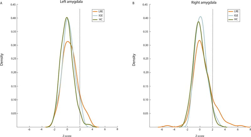
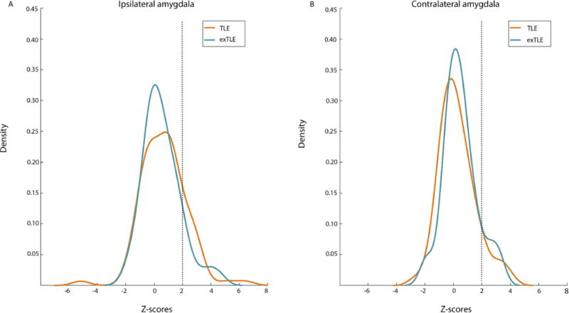
Results: AE was found in all groups including HCs; however, the rate of AE was higher in LRE (18.4%) than in IGE (5.9%) and HCs (6.4%). Patients with unilateral LRE were further evaluated to compare rates of concordant ipsilateral AE in TLE and exTLE, with the hypothesis that rates of ipsilateral AE would be higher in TLE. Although ipsilateral AE was higher in TLE (19.4%) than exTLE (10.5%), this difference was not significant. Furthermore, among the 25 patients with unilateral LRE and AE, 13 (52%) had either bilateral AE or AE contralateral to seizure onset.
Conclusion: Results suggest that AE, as defined with MRI volumetry, may represent an associated feature of nonlesional localization related epilepsy with limited seizure onset localization value.
Keywords: MRI; Morphometry; Nonlesional epilepsy; Temporal lobe epilepsy.
Copyright © 2017 Elsevier B.V. All rights reserved.
Figures
References
-
- Beh SJ, Cook MJ, D’Souza WJ. Isolated amygdala enlargement in temporal lobe epilepsy: A systematic review. Epilepsy & Behavior. 2016;60:33–41. - PubMed
-
- Fischl B, Salat DH, Busa E, Albert M, Dieterich M, Haselgrove C, Van Der Kouwe A, Killiany R, Kennedy D, Klaveness S, Montillo A. Whole brain segmentation: automated labeling of neuroanatomical structures in the human brain. Neuron. 2002;33(3):341–355. - PubMed
-
- Grimm O, Pohlack S, Cacciaglia R, Winkelmann T, Plichta MM, Demirakca T, Flor H. Amygdalar and hippocampal volume: A comparison between manual segmentation, Freesurfer and VBM. Journal of neuroscience methods. 2015;253:254–261. - PubMed
Publication types
MeSH terms
Grants and funding
LinkOut - more resources
Full Text Sources
Other Literature Sources
