. 2017 Mar 11;18(1):46.
doi: 10.1186/s12931-017-0529-6.

Combination of glycopyrronium and indacaterol inhibits carbachol-induced ERK5 signal in fibrotic processes

Affiliations

Combination of glycopyrronium and indacaterol inhibits carbachol-induced ERK5 signal in fibrotic processes

Yukiko Namba et al. Respir Res. .

Abstract

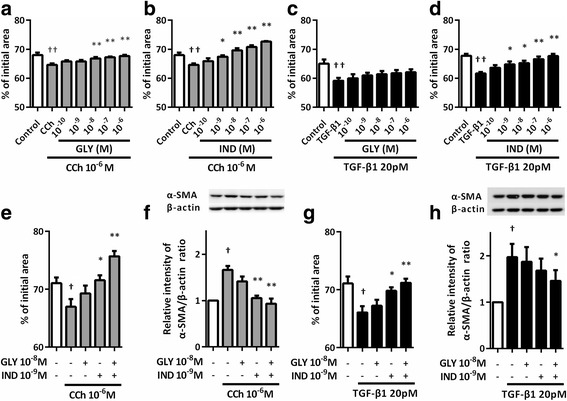
Background: Airway fibrosis is one of the pathological features of chronic obstructive pulmonary disease (COPD), and recent studies revealed that acetylcholine plays an important role in the development of airway remodeling by stimulating proliferation and collagen synthesis of lung fibroblasts. This study was designed to examine the effects of a long-acting muscarinic receptor antagonist (LAMA) glycopyrronium and a long-acting β2 adrenergic receptor agonist (LABA) indacaterol on acetylcholine-mediated fibrotic responses in lung fibroblasts.

Methods: After carbachol (CCh) or transforming growth factor-β1 (TGF-β1) exposure, the response to glycopyrronium and indacaterol was determined in vitro in fibroblasts isolated from mild-to-moderate COPD lung tissue. The ability of fibroblasts to mediate the contraction of collagen gels was assessed. The expression of α-smooth muscle actin (α-SMA) and the phosphorylation of extracellular-signal-regulated kinase 5 (ERK5) were determined by immunoblot. TGF-β1 was quantified by ELISA and acetylcholine was quantified by liquid chromatography tandem-mass spectrometry.

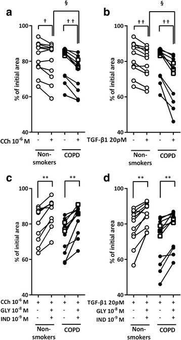
Results: CCh stimulated fibroblast-mediated collagen gel contraction and α-SMA expression and TGF-β1 release by fibroblasts. Blockade of autocrine TGF-β1 attenuated CCh-mediated fibrotic responses, while TGF-β1 did not stimulate acetylcholine release. Glycopyrronium plus indacaterol significantly attenuated CCh- and TGF-β1-mediated fibrotic responses through inhibition of ERK5 phosphorylation. Notably, the magnitudes of CCh- and TGF-β1-stimulated gel contraction, CCh-induced TGF-β1 release, and ERK5 phosphorylation were greater in fibroblasts isolated from COPD subjects than in those from non-smokers.

Conclusions: CCh induced TGF-β1 self-sustaining signaling loops by potentiating ERK5 signaling and promoted myofibroblast activity. This autocrine signaling mechanism may be an attractive therapeutic target to block the fibrotic response, which was modulated by the combination of glycopyrronium and indacaterol.

Keywords: Acetylcholine; Extracellular-signal-regulated kinase 5 (ERK5); Long-acting muscarinic receptor antagonist; Long-acting β2-adrenergic receptor agonist; Transforming growth factor-β1.


Figures

Fig. 1
Fig. 1
Effect of CCh, GLY or IND on collagen gel contraction and α-SMA expression in HFL-1 cells. Cells were grown to sub-confluence and deprived of serum for 24 h. Fibroblast-populated collagen gels were released into serum-free DMEM and measured the gel size daily by an image analyzer. a Gel size was measured in the presence of varying concentrations of CCh (b), GLY (e) or IND (f) on day3. Vertical axis, gel size expressed as % of initial size; Horizontal axis, conditions. All values represent the mean ± SEM in at least 3 separate experiments. HFL-1 cells were grown to sub-confluence in monolayer culture and stimulated with CCh at the indicated times or for 48 h at the indicated concentrations. After incubation, total protein was extracted and western blot analysis was performed with specific antibodies for α-SMA. Vertical axis: relative intensity of α-SMA/β-actin ratio. Horizontal axis: time (c) and concentration of CCh (d). All values represent the mean ± SEM in at least 3 separate experiments. *P < 0.05, **P < 0.01 compared with control
Fig. 2
Fig. 2
Effect of GLY and/or IND on CCh- or TGF-β1-augmented gel contraction and α-SMA expression in HFL1 cells. Sub-confluent fibroblasts were placed in serum-free (SF)-DMEM for 24 h. Cells were pre-treated with GLY and IND for 30 min, casted into collagen gels, and released into SF-DMEM containing various concentrations of GLY and/or IND with or without CCh (a, b, e) or TGF-β1 (c, d, g). Gel size was measured on day 3. Vertical axis: gel size expressed as a percentage of control. Horizontal axis: conditions. All values represent the mean ± SEM of three separate experiments, each performed in triplicate. For α-SMA assay, cells were grown to sub-confluence in monolayer culture and serum deprived for 24 h. Cells were then stimulated with CCh or TGF-β1 in the presence or absence of GLY and/or IND. After 48 h of incubation with CCh and 24 h of incubation with TGF-β1, cells were harvested for immunoblotting using a α-SMA antibody (f, h). Vertical axis, relative intensity of α-SMA/β-actin ratio; Horizontal axis, conditions. Representative immunoblots are shown at the top of each panel. All values represent the mean ± SEM in three different strains. P < 0.05, † † P < 0.01, compared with solvent control. *P < 0.05, **P < 0.01, compared with stimulus
Fig. 3
Fig. 3
CCh- or TGF-β1-augmented gel contraction was attenuated by GLY and IND in primary lung fibroblasts. After 24 h of SF-DMEM culture, lung fibroblasts from non-smokers or patients with COPD were pre-treated with GLY or IND for 30 min, casted into collagen gels and released into SF-DMEM in the presence or absence of CCh (a), TGF-β1 (b), with or without GLY/IND (c, d). Gel size was measured on day 3. Vertical axis: gel size expressed as a percentage of control. Horizontal axis: conditions. Each pair of symbols connected by a line represents data of an individual cell strain isolated from non-smokers or patients with COPD. Cells from non-smokers are indicated by open circles, cells from patients with COPD (GOLD I) by squares, and cells from patients with COPD (GOLD II) by closed circles. P < 0.05, † † P < 0.01, compared with control. *P < 0.05, **P < 0.01, compared with stimulus. § P < 0.05, compared with non-smokers and COPD lung fibroblasts in the same stimulus group
Fig. 4
Fig. 4
Effect of CCh-mediated endogenous TGF-β1 release in lung fibroblasts. Sub-confluent HFL1 cells (a) and primary lung fibroblasts (b) were cultured in SF-DMEM for 24 h in the presence or absence of CCh with or without GLY and/or IND. After 24 h of incubation, supernatants were harvested and used for TGF-β1 quantification. Vertical axis: TGF-β1 production expressed as amount per cells. Horizontal axis: conditions. Each pair of symbols connected by a line represents data of an individual cell strain isolated from non-smokers or patients with COPD. Cells from non-smokers are indicated by open circles, cells from patients with COPD (GOLD I) by squares, and cells from patients with COPD (GOLD II) by closed circles. c Sub-confluent HFL1 cells were cultured in SF-DMEM for 24 h. Cells were casted into collagen gels in the presence of CCh with or without SB431542, a selective inhibitor of the TGF-β1 receptor, ALK5. Gel size was measured on day 3. Vertical axis: gel size expressed as a percentage of control. Horizontal axis: conditions. All values represent the mean ± SEM of three separate experiments, each performed in triplicate. d HFL1 cells were grown to sub-confluence in monolayer culture and serum deprived for 24 h. Cells were stimulated with CCh in the presence or absence of SB431542. After 48 h of incubation, cells were harvested for the detection of α-SMA by immunoblotting. Vertical axis, relative intensity of α-SMA/β-actin ratio; Horizontal axis, conditions. All values represent the mean ± SEM of the three different strains. P < 0.05, compared with solvent control. *P < 0.05, compared with stimulus. § P < 0.05, compared with non-smokers and COPD lung fibroblasts in the same stimulus group
Fig. 5
Fig. 5
Determination of acetylcholine release by TGF-β1-stimulated lung fibroblasts by using LC-MS/MS. HFL1 and A549 cells were grown to sub-confluence in monolayer culture and serum deprived for 24 h. Cells were stimulated with TGF-β1 and harvested for the detection of ChAT by immunoblotting. Vertical axis, relative intensity of ChAT/β-actin ratio; Horizontal axis, conditions. Representative immunoblots are shown at the top of the panel. All values represent the mean ± SEM of the three different strains (a). HFL1 cells (b) and primary lung fibroblast from non-smokers (c) and patients with COPD (d) were grown to sub-confluence and cultured in SF-DMEM with or without TGF-β1 for 24 h. Supernatants were then harvested into a 1.5 ml tube and acetyl- and butyrylcholinesterase inhibitor (1 μM rivastigmine) was added to prevent acetylcholine degradation before storing at − 80 °C until assay. For the acetylcholine assay, the aliquots were thawed immediately before analysis and 5 μl of the samples was injected directly into the LC-MS/MS system. Data shown are representative chromatograms (m/z 86.910–87.910), performed in each group (n = 3). To obtain a standard report, acetylcholine was added to SF-DMEM. The arrow indicates the peak of acetylcholine (e). Vertical axis, signal intensity; Horizontal axis, retention time. *P < 0.05, **P < 0.01, compared with control
Fig. 6
Fig. 6
GLY and IND suppressed ERK5 phosphorylation in response to CCh or TGF-β1 in lung fibroblasts. Sub-confluent HFL1 cells were cultured in SF-DMEM with or without CCh (a) or TGF-β1 (b) for the indicated times. ERK5 phosphorylation was determined by immunoblotting. The bands with slower electrophoretic mobility corresponded to the activated kinase. Sub-confluent HFL1 cells (c, d) or primary fibroblasts (e-h) were serum deprived for 24 h followed by stimulation with or without TGF-β1 or CCh in the presence or absence of GLY and/or IND, SB431542 and ERK5 inhibitor (BIX02189) for 48 h. Cells were harvested and immunoblotted for ERK5. Each pair of symbols connected by a line represents data from an individual cell strain. Cells from non-smokers are indicated by open circles, cells from patients with COPD (GOLD I) by squares, and cells from patients with COPD (GOLD II) by closed circles. Vertical axis, the fold increase in activated kinase (p-ERK5) against total ERK5 over control or relative intensity of ERK5/β-actin ratio; Horizontal axis, times or conditions. All values represent the mean ± SEM. P < 0.05, compared with solvent control. *P < 0.05, **P < 0.01, compared with stimulus. § P < 0.05, compared with non-smokers and COPD lung fibroblasts in the same stimulus group
Fig. 7
Fig. 7
Inhibition of ERK5 resulted in the suppression of CCh- or TGF-β1-stimulated gel contraction and α-SMA expression in lung fibroblasts. Sub-confluent HFL1 cells were pre-treated with ERK5 inhibitor (BIX02189), ERK1/2 inhibitor (PD98059), and Rho kinase inhibitor (Y27632) for 30 min followed by being casted into collagen gels and treated with CCh or TGF-β1. Gel size was measured on day 3. Vertical axis: gel size expressed as a percentage of control. Horizontal axis: conditions. All values represent the mean ± SEM in three separate experiments, each performed in triplicate (a). Sub-confluent HFL1 cells were serum deprived for 24 h followed by stimulation with CCh or TGF-β1 for 48 h. Cells were then harvested for α-SMA immunoblotting. Vertical axis, relative intensity of α-SMA/β-actin ratio; Horizontal axis, conditions. All values represent the mean ± SEM in the three different strains (b). Lung fibroblasts from non-smokers and subjects with COPD were grown to sub-confluence and pre-treated with BIX02189 for 30 min followed by being casted into collagen gels and released into SF-DMEM with CCh (c) or TGF-β1 (d) in the presence or absence of BIX02189. Gel size was measured on day 3. Vertical axis: gel size expressed as a percentage of control. Horizontal axis: conditions. Each pair of symbols connected by a line represented data from an individual cell strain and the mean of three separate experiments. Cells from non-smokers are indicated by open circles, cells from patients with COPD (GOLD I) by squares, and cells from patients with COPD (GOLD II) by closed circles. P < 0.05, †† P < 0.01, compared with solvent control. *P < 0.05, **P < 0.01, compared with stimulus. § P < 0.05, compared with non-smokers and COPD lung fibroblasts in the same stimulus group
Fig. 8
Fig. 8
Schematic illustration of CCh-induced endogenous TGF-β1 signaling loop in COPD lung fibroblasts. Carbachol (CCh) induces ERK5 activation directly or indirectly through stimulating endogenous transforming growth factor-β1 (TGF-β1) release. Glycopyrronium (GLY) directly blocks CCh binding to its receptor, while indacaterol (IND) interrupts CCh signaling pathway or TGF-β1-induced ERK5 activation. Through these mechanisms, GLY plus IND blocked CCh- or TGF-β1-augmented collagen gel contraction and α-SMA expression

Similar articles

Cited by

References

    1. Berankova K, Uhlik J, Honkova L, Pohunek P. Structural changes in the bronchial mucosa of young children at risk of developing asthma. Pediatr Allergy Immunol. 2014;25:136–142. doi: 10.1111/pai.12119. - DOI - PubMed
    1. Bhatt SP, Soler X, Wang X, Murray S, Anzueto AR, Beaty TH, Boriek AM, Casaburi R, Criner GJ, Diaz AA, et al. Association Between Functional Small Airways Disease and FEV Decline in COPD. Am J Respir Crit Care Med. 2016. - PMC - PubMed
    1. Hogg JC, Chu F, Utokaparch S, Woods R, Elliott WM, Buzatu L, Cherniack RM, Rogers RM, Sciurba FC, Coxson HO, Pare PD. The nature of small-airway obstruction in chronic obstructive pulmonary disease. N Engl J Med. 2004;350:2645–2653. doi: 10.1056/NEJMoa032158. - DOI - PubMed
    1. Rennard SI, Wachenfeldt K. Rationale and emerging approaches for targeting lung repair and regeneration in the treatment of chronic obstructive pulmonary disease. Proc Am Thorac Soc. 2011;8:368–375. doi: 10.1513/pats.201102-019RM. - DOI - PubMed
    1. Takizawa H, Tanaka M, Takami K, Ohtoshi T, Ito K, Satoh M, Okada Y, Yamasawa F, Nakahara K, Umeda A. Increased expression of transforming growth factor-beta1 in small airway epithelium from tobacco smokers and patients with chronic obstructive pulmonary disease (COPD) Am J Respir Crit Care Med. 2001;163:1476–1483. doi: 10.1164/ajrccm.163.6.9908135. - DOI - PubMed

Publication types

MeSH terms