Review
. 2017 May;187(5):940-953.
doi: 10.1016/j.ajpath.2017.01.010. Epub 2017 Mar 9.

Hypoxia-Mediated Mechanisms Associated with Antiangiogenic Treatment Resistance in Glioblastomas

Affiliations
Review

Hypoxia-Mediated Mechanisms Associated with Antiangiogenic Treatment Resistance in Glioblastomas

Sean Mahase et al. Am J Pathol. 2017 May.

Abstract

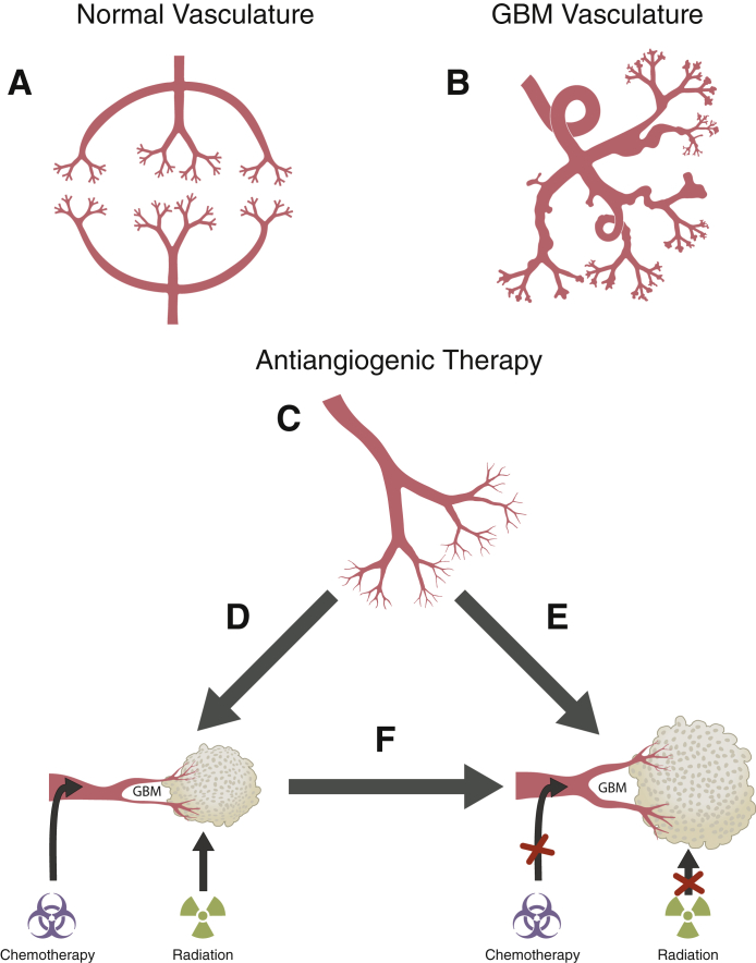
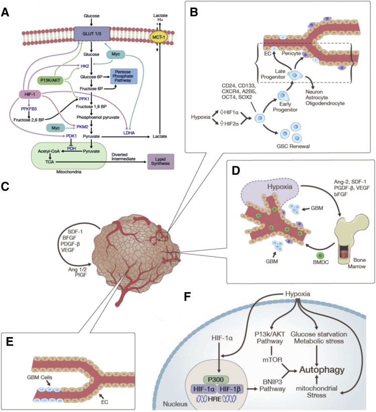
Glioblastomas (GBMs) are malignant tumors characterized by their vascularity and invasive capabilities. Antiangiogenic therapy (AAT) is a treatment option that targets GBM-associated vasculature to mitigate the growth of GBMs. However, AAT demonstrates transient effects because many patients eventually develop resistance to this treatment. Several recent studies attempt to explain the molecular and biochemical basis of resistance to AAT in GBM patients. Experimental investigations suggest that the induction of extensive intratumoral hypoxia plays a key role in GBM escape from AAT. In this review, we examine AAT resistance in GBMs, with an emphasis on six potential hypoxia-mediated mechanisms: enhanced invasion and migration, including increased expression of matrix metalloproteinases and activation of the c-MET tyrosine kinase pathway; shifts in cellular metabolism, including up-regulation of hypoxia inducible factor-1α's downstream processes and the Warburg effect; induction of autophagy; augmentation of GBM stem cell self-renewal; possible implications of GBM-endothelial cell transdifferentiation; and vasoformative responses, including vasculogenesis, alternative angiogenic pathways, and vascular mimicry. Juxtaposing recent studies on well-established resistance pathways with that of emerging mechanisms highlights the overall complexity of GBM treatment resistance while also providing direction for further investigation.


Figures

Figure 1
Figure 1
Twenty-two-year-old female, previously treated for an anaplastic astrocytoma at New York University Langone Medical Center, started on bevacizumab (BEV) 6 months after the initial diagnosis for a progressive recurrent tumor. Baseline imaging before BEV treatment: axial flair (A), axial view after contrast (B), and DSC T2*cerebral blood volume parametric maps (C) showing a large, recurrent, heterogeneously enhancing glioblastoma with surrounding edema and mass effect, as well as markedly increased blood volume. Four weeks after BEV treatment: axial flair (D), axial view after contrast (E), and dynamic susceptibility contrast (DSC) T2*cerebral blood volume parametric maps (F) showing slight improvement in the size and degree of enhancement of the large heterogenous tumor, with slight improvement in the swelling and edema. Remarkably, cerebral blood volume maps show reduction in tumor blood volume, suggestive of typical initial response seen with antiangiogenic therapy.
Figure 2
Figure 2
A series of T1-weighted images after contrast depict the clinical course of a 41-year-old man with a large recurrence of glioblastoma in the right cerebral hemisphere who received 17 administrations of bevacizumab (BEV) over 36 months. A: Baseline imaging before starting BEV shows heterogenous areas of enhancement. B–D: Six (B), 30 (C), and 38 (D) weeks of BEV therapy shows progressively decreasing size of the contrast-enhancing lesion with improvement in swelling and mass effect. E: Six weeks after stopping BEV the image shows recurrence of the enhancing lesion. F: Twelve weeks after stopping BEV the image shows dramatic increase in enhancement. The patient died 20 weeks after stopping BEV therapy because of progressive increase in size of the tumor.
Figure 3
Figure 3
Proposed effect of antiangiogenic therapy on glioblastoma (GBM) vasculature. A: Normal brain vasculature. B: GBM vasculature: aberrant vascular architecture with dilated, highly permeable blood vessels and multiple blind loops. C: After BEV treatment: BEV-mediated normalization of glioma vasculature is denoted by decreased vessel permeability and a reduced number of large- and mid-sized vessels. D: Theory 1: anti-VEGF treatment restores perfusion, promoting improved chemotherapy delivery and radiation therapy efficacy. E: Theory 2: structural changes entailing vessel normalization do not result in completely functional or normal vasculature. F: Theory 3: At some point after treatment, the anti-VEGF–induced vasculature normalization is eventually lost for reasons still to be (further) elucidated, and the tumor becomes resistant to additional antiangiogenic therapy. BEV, bevacizumab; VEGF, vascular endothelial growth factor.
Figure 4
Figure 4
Hypoxia-mediated mechanisms of resistance to therapeutic modalities (see Figure 2 and Tables 1 and 2 for illustrations of enhanced migration and invasion): shifts in cellular metabolism (A); GSCs and GBM-EC transdifferentiation (B); vasoformative responses entailing alternate angiogenic pathways (C), vasculogenesis (D), and vascular mimicry (E); and autophagy (F). Ang, angiopoietin; bFGF, basic fibroblast growth factor; BMDC, bone marrow-derived cell; BNIP, Bcl/E1B 19kD interacting protein; BP, bisphosphate; CoA, coenzyme A; EC, endothelial cell; GBM, glioblastoma; GLUT, glucose transporter; GSC, glioblastoma stem cell; HIF, hypoxia-inducible factor; HK2, hexokinase-2; HRE, hypoxia-response element; LDHA, lactate dehydrogenase A; MCT, monocarboxylate transporter; mTOR, mechanistic target of rapamycin; OCT4, octamer-binding transcription factor 4; P, phosphate; PDGF, platelet-derived growth factor; PDH, pyruvate dehydrogenase; PDK, pyruvate dehydrogenase kinase; PFKFB3, 6-phosphofructo-2-kinase/fructose-2,6-biphosphatase 3; PI3K, phosphinositol-3-kinase; PKM2, pyruvate kinase muscle isozyme M2; PlGF, placental growth factor; SDF, stromal cell-derived factor; Sox2, sex determining region Y-box 2; TCA, tricarboxylic acid; VEGF, vascular endothelial growth factor.

References

    1. Woehrer A., Bauchet L., Barnholtz-Sloan J.S. Glioblastoma survival: has it improved? Evidence from population-based studies. Curr Opin Neurol. 2014;27:666–674. - PubMed
    1. Wen P.Y., Kesari S. Malignant gliomas in adults. N Engl J Med. 2008;359:492–507. - PubMed
    1. Fischer I., Gagner J.P., Law M., Newcomb E.W., Zagzag D. Angiogenesis in gliomas: biology and molecular pathophysiology. Brain Pathol. 2005;15:297–310. - PMC - PubMed
    1. Gilbert M.R., Dignam J.J., Armstrong T.S., Wefel J.S., Blumenthal D.T., Vogelbaum M.A., Colman H., Chakravarti A., Pugh S., Won M., Jeraj R., Brown P.D., Jaeckle K.A., Schiff D., Stieber V.W., Brachman D.G., Werner-Wasik M., Tremont-Lukats I.W., Sulman E.P., Aldape K.D., Curran W.J., Jr., Mehta M.P. A randomized trial of bevacizumab for newly diagnosed glioblastoma. N Engl J Med. 2014;370:699–708. - PMC - PubMed
    1. Jain R.K., di Tomaso E., Duda D.G., Loeffler J.S., Sorensen A.G., Batchelor T.T. Angiogenesis in brain tumours. Nat Rev Neurosci. 2007;8:610–622. - PubMed

MeSH terms