. 2017 May 15:152:371-380.
doi: 10.1016/j.neuroimage.2017.02.074. Epub 2017 Mar 8.

Functional connectivity and activity of white matter in somatosensory pathways under tactile stimulations

Affiliations

Functional connectivity and activity of white matter in somatosensory pathways under tactile stimulations

Xi Wu et al. Neuroimage. .

Abstract

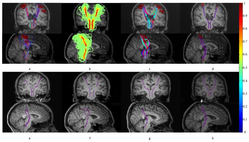
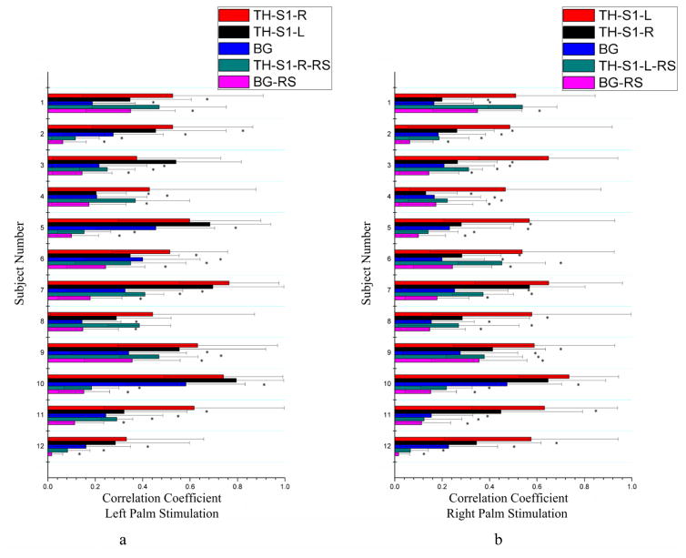
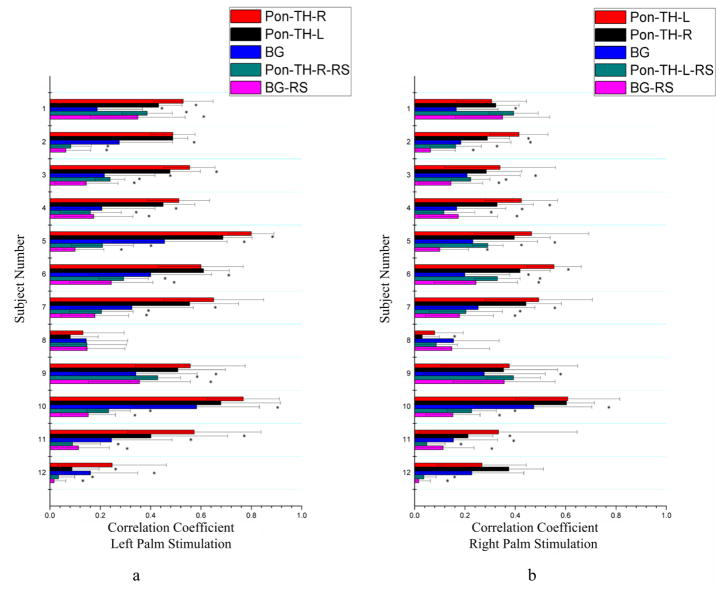
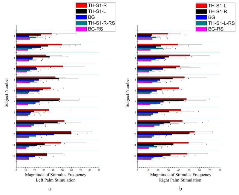
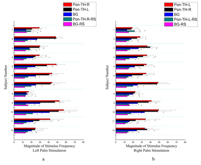
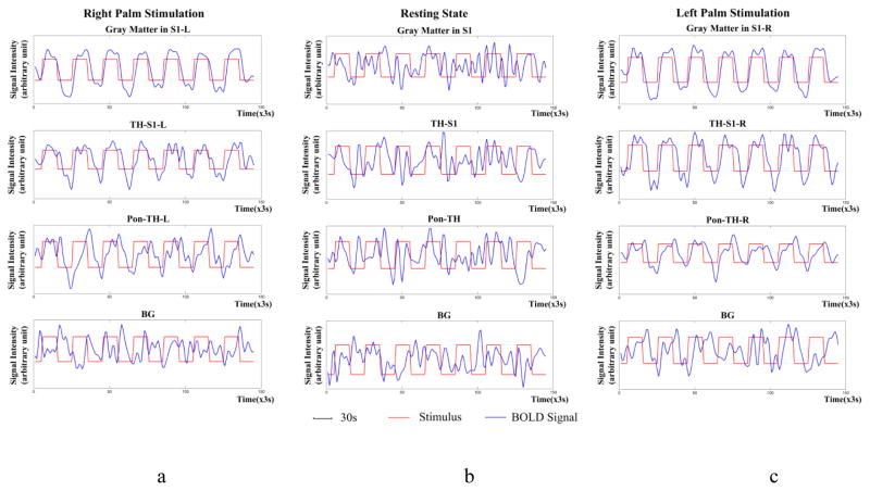
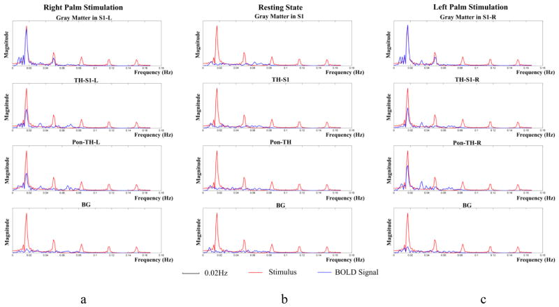
Functional MRI has proven to be effective in detecting neural activity in brain cortices on the basis of blood oxygenation level dependent (BOLD) contrast, but has relatively poor sensitivity for detecting neural activity in white matter. To demonstrate that BOLD signals in white matter are detectable and contain information on neural activity, we stimulated the somatosensory system and examined distributions of BOLD signals in related white matter pathways. The temporal correlation profiles and frequency contents of BOLD signals were compared between stimulation and resting conditions, and between relevant white matter fibers and background regions, as well as between left and right side stimulations. Quantitative analyses show that, overall, MR signals from white matter fiber bundles in the somatosensory system exhibited significantly greater temporal correlations with the primary sensory cortex and greater signal power during tactile stimulations than in a resting state, and were stronger than corresponding measurements for background white matter both during stimulations and in a resting state. The temporal correlation and signal power under stimulation were found to be twice those observed from the same bundle in a resting state, and bore clear relations with the side of stimuli. These indicate that BOLD signals in white matter fibers encode neural activity related to their functional roles connecting cortical volumes, which are detectable with appropriate methods.

Keywords: Diffusion MRI; Functional MRI; Sensory stimulation; White matter.


Figures

Figure 1
Figure 1
Tracking and skeletonization of the projection pathways. (a) Probabilistic tracking of fibers from thalamus to S1 (and pons). (b) Probability density of fibers connecting thalamus and S1 (and pons). (c) Streamlines generated by backtracking from S1 (and pons) to thalamus. (d) Skeletons of fiber bundles connecting thalamus and S1 (and pons). First and second rows are coronal and sagittal views respectively. (e–h) Representative skeletons from four other subjects shown in coronal and sagittal views. Note that the colorbar on the far right is for panel (b).
Figure 2
Figure 2
Comparisons of mean correlations between signals along the bundle skeleton connecting the thalamus to S1 and S1 for tactile stimulation and resting state conditions. Background (BG) included 1000 random white matter voxels outside the fiber bundles connecting the thalamus and S1 (and pons). Left (a) and right (b) columns are for left and right palm stimulations respectively. *denotes p<0.05. See text for explanations of the abbreviations.
Figure 3
Figure 3
Comparisons of mean correlations between signals along the bundle skeleton connecting the thalamus to the pons and the pons for stimulation and resting state conditions. BG area included 1000 random white matter voxels outside the fiber bundles connecting the thalamus and pons (and S1). Left (a) and right (b) columns are for left and right palm stimulations respectively. *denotes p<0.05. See text for explanations of the abbreviations.
Figure 4
Figure 4
Comparisons of mean signal magnitude at stimulus frequency along the bundle skeleton connecting the thalamus and the S1 area between tactile stimulation and resting state conditions. Left (a) and right (b) columns are for left and right palm stimulations respectively. *denotes p<0.05. See text for explanations of the abbreviations.
Figure 5
Figure 5
Comparisons of mean signal magnitude at stimulus frequency along the bundle skeleton connecting the thalamus and the pons area between tactile stimulation and resting state conditions. Left (a) and right (b) columns are for left and right palm stimulations respectively. *denotes p<0.05. See text for explanations of the abbreviations.
Figure 6
Figure 6
Temporal variations of BOLD signals averaged across the twelve subjects studied. From top to bottom rows are mean BOLD signals in S1, along the bundle skeleton connecting thalamus and S1, along the bundle skeleton connecting thalamus and pons, and background respectively. From left to right columns are right palm stimulations (a), resting state condition (b) and left palm stimulations (c) respectively. The signal intensity is in arbitrary unit and red and blue curves represent stimulus and BOLD signal respectively. Note that, unlike stimulation conditions, BOLD signals in the resting state contain no stimulus-related periodicity.
Figure 7
Figure 7
Power spectra of mean BOLD signals in Figure 6.

References

    1. Astafiev SV, Shulman GL, Metcalf NV, Rengachary J, MacDonald CL, Harrington DL, Maruta J, Shimony JS, Ghajar J, Diwakar M, Huang MX, Lee RR, Corbetta M. Abnormal White Matter Blood-Oxygen-Level-Dependent Signals in Chronic Mild Traumatic Brain Injury. Neurotrauma. 2015;32:1254–1271. - PMC - PubMed
    1. Beckmann CF, DeLuca M, Devlin JT, Smith SM. Investigations into resting-state connectivity using independent component analysis. Philos Trans Royal Soc Lond B Biol Sci. 2005;360:1001–1013. - PMC - PubMed
    1. Biswal B, Yetkin FZ, Haughton VM, Hyde JS. Functional connectivity in the motor cortex of resting human brain using echo-planar MRI. Magn Reson Med. 1995;34:537–541. - PubMed
    1. Bodurka J, Ye F, Petridou N, Murphy K, Bandettini P. Mapping the MRI voxel volume in which thermal noise matches physiological noise-Implications for fMRI. Neuroimage. 2007;34:542–549. - PMC - PubMed
    1. Bullmore E, Fadili J, Maxim V, Sendur L, Whitcher B, Suckling J, Brammer M, Breakspear M. Wavelets and functional magnetic resonance imaging of the human brain. Neuroimage. 2004;23:234–249. - PubMed

Publication types

LinkOut - more resources