

Engineering epithelial-stromal interactions in vitro for toxicology assessment
- PMID: 28285100
- PMCID: PMC5985517
- DOI: 10.1016/j.tox.2017.03.007
Engineering epithelial-stromal interactions in vitro for toxicology assessment
Abstract
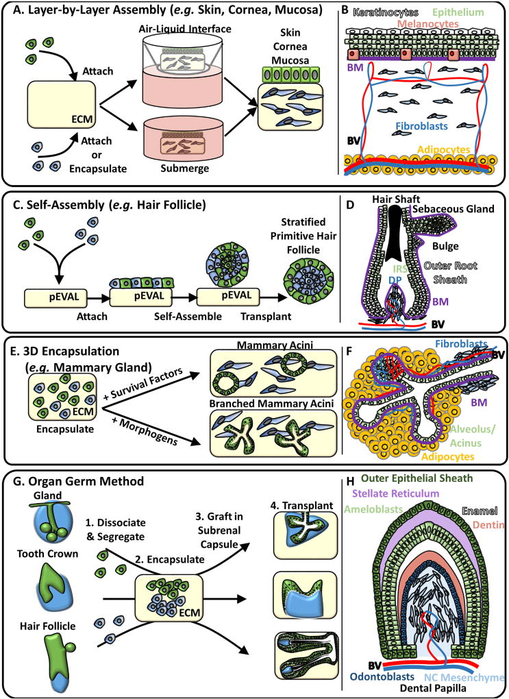
Crosstalk between epithelial and stromal cells drives the morphogenesis of ectodermal organs during development and promotes normal mature adult epithelial tissue homeostasis. Epithelial-stromal interactions (ESIs) have historically been examined using mammalian models and ex vivo tissue recombination. Although these approaches have elucidated signaling mechanisms underlying embryonic morphogenesis processes and adult mammalian epithelial tissue function, they are limited by the availability of tissue, low throughput, and human developmental or physiological relevance. In this review, we describe how bioengineered ESIs, using either human stem cells or co-cultures of human primary epithelial and stromal cells, have enabled the development of human in vitro epithelial tissue models that recapitulate the architecture, phenotype, and function of adult human epithelial tissues. We discuss how the strategies used to engineer mature epithelial tissue models in vitro could be extrapolated to instruct the design of organotypic culture models that can recapitulate the structure of embryonic ectodermal tissues and enable the in vitro assessment of events critical to organ/tissue morphogenesis. Given the importance of ESIs towards normal epithelial tissue development and function, such models present a unique opportunity for toxicological screening assays to incorporate ESIs to assess the impact of chemicals on mature and developing epidermal tissues.
Keywords: Bioengineering; Development; Epithelial cells; Epithelial-stromal interactions; Morphogenesis; Multipotent stromal cells; Organotypic; Palate fusion; Stromal cells.
Published by Elsevier B.V.
Conflict of interest statement
The authors have declared that no competing interests exist
Figures

Similar articles
-
Engineering human cell spheroids to model embryonic tissue fusion in vitro.PLoS One. 2017 Sep 12;12(9):e0184155. doi: 10.1371/journal.pone.0184155. eCollection 2017. PLoS One. 2017. PMID: 28898253 Free PMC article.
-
A complex 3D human tissue culture system based on mammary stromal cells and silk scaffolds for modeling breast morphogenesis and function.Biomaterials. 2010 May;31(14):3920-9. doi: 10.1016/j.biomaterials.2010.01.118. Epub 2010 Feb 24. Biomaterials. 2010. PMID: 20185172 Free PMC article.
-
Stromal-epithelial cell interactions and alteration of branching morphogenesis in macromastic mammary glands.J Cell Mol Med. 2014 Jul;18(7):1257-66. doi: 10.1111/jcmm.12275. Epub 2014 Apr 10. J Cell Mol Med. 2014. PMID: 24720804 Free PMC article.
-
Concise review: can the intrinsic power of branching morphogenesis be used for engineering epithelial tissues and organs?Stem Cells Transl Med. 2013 Dec;2(12):993-1000. doi: 10.5966/sctm.2013-0076. Epub 2013 Nov 4. Stem Cells Transl Med. 2013. PMID: 24191267 Free PMC article. Review.
-
Biomaterials and bioengineering to guide tissue morphogenesis in epithelial organoids.Front Bioeng Biotechnol. 2022 Nov 17;10:1038277. doi: 10.3389/fbioe.2022.1038277. eCollection 2022. Front Bioeng Biotechnol. 2022. PMID: 36466337 Free PMC article. Review.
Cited by
-
Organoid-based tissue engineering for advanced tissue repair and reconstruction.Mater Today Bio. 2025 Jul 15;33:102093. doi: 10.1016/j.mtbio.2025.102093. eCollection 2025 Aug. Mater Today Bio. 2025. PMID: 40727081 Free PMC article. Review.
-
Biofabrication of a Tubular Model of Human Urothelial Mucosa Using Human Wharton Jelly Mesenchymal Stromal Cells.Polymers (Basel). 2021 May 13;13(10):1568. doi: 10.3390/polym13101568. Polymers (Basel). 2021. PMID: 34068343 Free PMC article.
-
Engineering human cell spheroids to model embryonic tissue fusion in vitro.PLoS One. 2017 Sep 12;12(9):e0184155. doi: 10.1371/journal.pone.0184155. eCollection 2017. PLoS One. 2017. PMID: 28898253 Free PMC article.
-
Scaffold-based developmental tissue engineering strategies for ectodermal organ regeneration.Mater Today Bio. 2021 Mar 6;10:100107. doi: 10.1016/j.mtbio.2021.100107. eCollection 2021 Mar. Mater Today Bio. 2021. PMID: 33889838 Free PMC article. Review.
References
-
- Abbott BD. Experimental models for the study of oral clefts. In: Wyszynski DF, editor. Cleft Lip and Palate: From Origin to Treatment. Oxford University Press; Cary, NC: 2002. pp. 193–202.
-
- Abbott BD. The etiology of cleft palate: a 50-year search for mechanistic and molecular understanding. Birth Defects Res. B. 2010;89:266–274. - PubMed
-
- Arrighi S. The urothelium: anatomy, review of the literature, perspectives for veterinary medicine. Ann. Anat. Anatom. Anz. 2015;198:73–82. - PubMed
Publication types
MeSH terms
Grants and funding
LinkOut - more resources
Full Text Sources
Other Literature Sources
