. 2017 May;309(4):285-303.
doi: 10.1007/s00403-017-1729-0. Epub 2017 Mar 11.

Comparison of protective effect of ascorbic acid on redox and endocannabinoid systems interactions in in vitro cultured human skin fibroblasts exposed to UV radiation and hydrogen peroxide

Affiliations

Comparison of protective effect of ascorbic acid on redox and endocannabinoid systems interactions in in vitro cultured human skin fibroblasts exposed to UV radiation and hydrogen peroxide

Agnieszka Gęgotek et al. Arch Dermatol Res. 2017 May.

Abstract

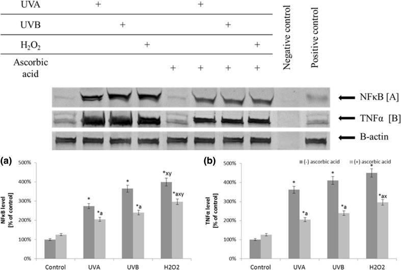
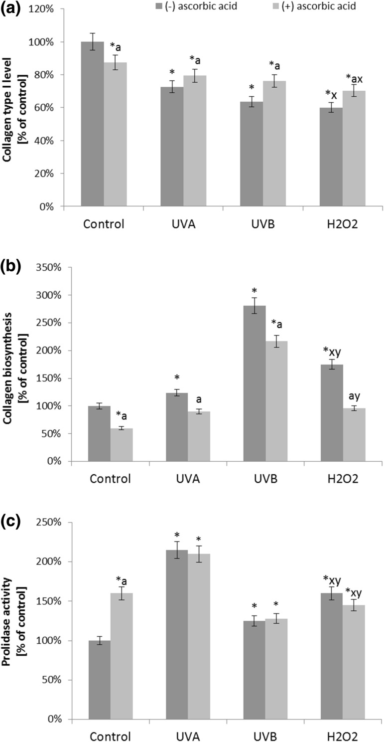
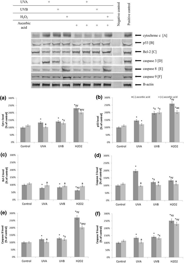
The mechanisms of biological activity of commonly used natural compounds are constantly examined. Therefore, the aim of this study was to compare ascorbic acid efficacy in counteracting the consequences of UV and hydrogen peroxide treatment on lipid mediators and their regulative action on antioxidant abilities. Skin fibroblasts exposed to UVA and UVB irradiation, treated with hydrogen peroxide and ascorbic acid. The redox system was estimated through reactive oxygen species (ROS) generation (electron spin resonance spectrometer) and antioxidants level/activity (HPLC/spectrometry) which activity was evaluated by the level of phospholipid metabolites: 4-hydroxynonenal, malondialdehyde, 8-isoprostanes and endocannabinoids (GC/LC-MS) in the human skin fibroblasts. Protein and DNA oxidative modifications were also determined (LC). The expression of nuclear factor erythroid 2-related factor 2 (Nrf2), its activators and inhibitors as well as pro/anti-apoptotic proteins and endocannabinoid receptors was examined (Western blot) and collagen metabolism was evaluated by collagen biosynthesis and prolidase activity (spectrometry). UVA and UVB irradiation and hydrogen peroxide treatment enhanced activity of xanthine and NADPH oxidases resulting in ROS generation as well as diminution of antioxidant phospholipid protection (glutathione peroxidase-glutathione-vitamin E), what led to increased lipid peroxidation and decreased endocannabinoids level. Dysregulation of cannabinoid receptors expression and environment of transcription factor Nrf2 caused apoptosis induction. Ascorbic acid partially prevented ROS generation, antioxidant capacity diminution and endocannabinoid systems disturbances but only slightly protected macromolecules such as phospholipid, protein and DNA against oxidative modifications. However, ascorbic acid significantly prevented decrease in collagen type I biosynthesis. Ascorbic acid in similar degree prevents UV (UVA and UVB) and hydrogen peroxide-dependent redox imbalance. However, this antioxidant cannot efficiently protect cellular macromolecules and avert metabolic dysregulation leading to apoptosis.

Keywords: Ascorbic acid; Endocannabinoid system; Fibroblasts; Hydrogen peroxide; Nrf2; UV radiation.


Conflict of interest statement

Conflict of interest

The authors declare that they have no conflict of interest.

Figures

Fig. 1
Fig. 1
The comparison of cell viability, ROS generation, and 4-HNE level in fibroblasts after exposure to UVA (20 J/cm2), UVB radiation (200 mJ/cm2), H2O2 (200 µM), and ascorbic acid (100 µM) expressed as a percentage of the value of the control cells. Mean values ± SD of three independent experiments are presented. Asterisk statistically significant differences vs. control group, p < 0.05; a statistically significant differences vs. group without ascorbic acid, p < 0.05; x statistically significant differences vs. UVA group, p < 0.05; y statistically significant differences vs. UVB group, p < 0.05
Fig. 2
Fig. 2
The level of transcription factor NFκB and its target protein—TNFα in fibroblasts after exposure to UVA (20 J/cm2), UVB radiation (200 mJ/cm2), H2O2 (200 µM), and ascorbic acid (100 µM) expressed as a percentage of the value of the control cells. Mean values ± SD of three independent experiments are presented. Asterisk statistically significant differences vs. control group, p < 0.05; a statistically significant differences vs. group without ascorbic acid, p < 0.05; x statistically significant differences vs. UVA group, p < 0.05; y statistically significant differences vs. UVB group, p < 0.05
Fig. 3
Fig. 3
The xanthine oxidase (a) and NADPH oxidase (b) activity, and superoxide anion generation (c) in fibroblasts after exposure to UVA (20 J/cm2), UVB radiation (200 mJ/cm2), H2O2 (200 µM), and ascorbic acid (100 µM) expressed as a percentage of the value of the control cells. Mean values ± SD of three independent experiments are presented. Asterisk statistically significant differences vs. control group, p < 0.05; a statistically significant differences vs. group without ascorbic acid, p < 0.05; x statistically significant differences vs. UVA group, p < 0.05; y statistically significant differences vs. UVB group, p < 0.05
Fig. 4
Fig. 4
The level of Nrf2 (a), its phosphorylated form (pSer40) (b), its inhibitors: Keap1, Bach1 (c, d), and activators: KAP1, p21, p62 (e, f, g), and HO-1 (h) in fibroblasts after exposure to UVA (20 J/cm2), UVB radiation (200 mJ/cm2), H2O2 (200 µM), and ascorbic acid (100 µM) expressed as a percentage of the value of the control cells. Mean values ± SD of three independent experiments are presented. Asterisk statistically significant differences vs. control group, p < 0.05; a statistically significant differences vs. group without ascorbic acid, p < 0.05; x statistically significant differences vs. UVA group, p < 0.05; y statistically significant differences vs. UVB group, p < 0.05
Fig. 5
Fig. 5
The cytoplasmic and nucleus level of Nrf2 in fibroblasts control cells and after exposure of UVA (20 J/cm2), UVB radiation (200 mJ/cm2), H2O2 (200 µM), and ascorbic acid (100 µM) (blue nucleus; red Nrf2)
Fig. 6
Fig. 6
The level of endocannabinoids (AEA and 2-AG) (a, b) and the expression of endocannabinoids receptors (CB1, CB2, VR1, GPR55) (cf) in fibroblasts after exposure to UVA (20 J/cm2), UVB radiation (200 mJ/cm2), H2O2 (200 µM), and ascorbic acid (100 µM) expressed as a percentage of the value of the control cells. Mean values ± SD of three independent experiments are presented. *Statistically significant differences vs. control group, p < 0.05; a statistically significant differences vs. group without ascorbic acid, p < 0.05; x statistically significant differences vs. UVA group, p < 0.05; y statistically significant differences vs. UVB group, p < 0.05;
Fig. 7
Fig. 7
The level of collagen type I expression (a), its biosynthesis (b) and prolidase activity (c) in fibroblasts after exposure to UVA (20 J/cm2), UVB radiation (200 mJ/cm2), H2O2 (200 µM), and ascorbic acid (100 µM) expressed as a percentage of the value of the control cells. Mean values ± SD of three independent experiments are presented. Asterisk statistically significant differences vs. control group, p < 0.05; a statistically significant differences vs. group without ascorbic acid, p < 0.05; x statistically significant differences vs. UVA group, p < 0.05; y statistically significant differences vs. UVB group, p < 0.05
Fig. 8
Fig. 8
The level of pro- and anti-apoptotic proteins [cytochrome c (a), p53 (b), Bcl-2 (c)], and executive caspases [3, 8, and 9 (df)] in fibroblasts after exposure to UVA (20 J/cm2), UVB radiation (200 mJ/cm2), H2O2 (200 µM), and ascorbic acid (100 µM) expressed as a percentage of the value of the control cells. Mean values ± SD of three independent experiments are presented. Asterisk statistically significant differences vs. control group, p < 0.05; a statistically significant differences vs. group without ascorbic acid, p < 0.05; x statistically significant differences vs. UVA group, p < 0.05; y statistically significant differences vs. UVB group, p < 0.05
Fig. 9
Fig. 9
A scheme summarizing the ascorbic acid effect on ROS generation, lipid mediators and its consequences for the redox homeostasis in fibroblasts following exposure to UVA and UVB radiation, as well as H2O2 treatment

References

    1. Birch-Machin MA, Swalwell H. How mitochondria record the effects of UV exposure and oxidative stress using human skin as a model tissue. Mutagenesis. 2009;25:101–107. doi: 10.1093/mutage/gep061. - DOI - PubMed
    1. Hakozaki T, Date A, Yoshii T, Toyokuni S, Yasui H, Sakurai H. Visualization and characterization of UVB-induced reactive oxygen species in a human skin equivalent model. Arch Dermatol Res. 2008;300:51–56. doi: 10.1007/s00403-007-0804-3. - DOI - PubMed
    1. Cadet J, Berger M, Douki T, Morin B, Raoul S, Ravanat JL, et al. Effects of UV and visible radiation on DNA-final base damage. Biol Chem. 1997;378:1275–1286. - PubMed
    1. Dash R, Acharya C, Bindu PC, Kundu SC. Antioxidant potential of silk protein sericin against hydrogen peroxide-induced oxidative stress in skin fibroblasts. BMB Rep. 2008;41:236–241. doi: 10.5483/BMBRep.2008.41.3.236. - DOI - PubMed
    1. Oyewole A, Birket MJ, Levett D, Anderson M, Swalwell H, Birch-Machin MA. British society for investigative dermatology annual meeting. New Jersey: Wiley-Blackwell Publishing Ltd; 2009. pp. 924–925.

MeSH terms