Comparative Study
. 2017 Mar 13;10(1):113.
doi: 10.1186/s13071-017-2050-7.

Canine leishmaniosis caused by Leishmania major and Leishmania tropica: comparative findings and serology

Affiliations
Comparative Study

Canine leishmaniosis caused by Leishmania major and Leishmania tropica: comparative findings and serology

Gad Baneth et al. Parasit Vectors. .

Abstract

Background: Infection and clinical disease associated with Leishmania major and Leishmania tropica, two common agents of human cutaneous leishmaniosis, have rarely been reported in dogs. This study describes dogs infected with these Leishmania spp. prevalent in the Middle East and North Africa, and compares the serological response of dogs infected with Leishmania infantum, L. major or L. tropica to whole promastigote antigen enzyme-linked immunosorbent assay (ELISA) of each species and to rK39 dipstick.

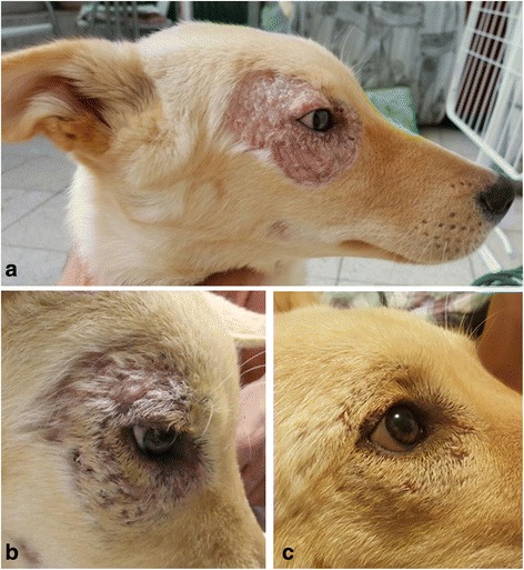
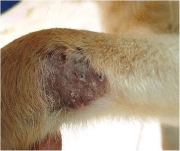
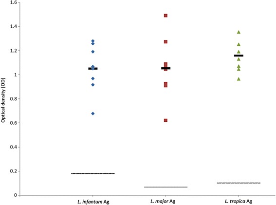
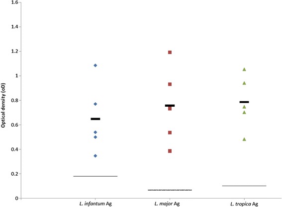
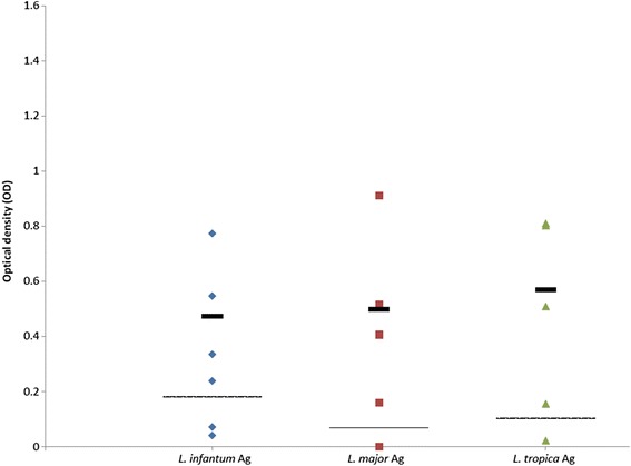
Results: Leishmania major infection in a 5-month-old male dog was associated with alopecic and ulcerative periocular and limb skin lesions which responded to allopurinol treatment. Infection was detected by skin and blood polymerase chain reaction (PCR) and confirmed by DNA sequencing but the dog was seronegative. Leishmania tropica infection was detected in a 3-month-old female dog co-infected with Babesia vogeli and Anaplasma platys and with no skin lesions. PCR and DNA sequencing of the blood and parasite culture were positive for L. tropica. Sera from 11 dogs infected with L. infantum, L. major or L. tropica were reactive with all three Leishmania spp. antigens except for sera from a dog with L. major infection. No significant differences were found between reactivity of dog sera to the antigen of the infecting species, or to the other Leishmania spp. antigens. Sera from dogs infected with L. infantum and L. tropica were positive with the rK39 antigen kit, while dogs with L. major infection were seronegative.

Conclusions: Skin lesions in L. major infected dogs from this study and previous reports (n = 2) were ulcerative and located on the muzzle, feet and foot pads and not associated with generalized lymphadenomegaly and splenomegaly. In previous L. tropica infections, skin lesions were proliferative mucocutaneous in young dogs (n = 2), or associated with widespread dermatitis, lymphadenomegaly and splenomegaly in older dogs with similarity to L. infantum infection (n = 2). This study suggests that ELISA serology with whole promastigote antigen is not distinctive between L. infantum, L. major and L. tropica canine infections and that some L. major infections are not seropositive. PCR with DNA sequencing should be used to discriminate between canine infections with these three species.

Keywords: Canine leishmaniosis; Co-infections; Cutaneous leishmaniosis; Israel; Leishmania major; Leishmania tropica.


Figures

Fig. 1
Fig. 1
Cutaneous periocular Leishmania major skin lesion in dog case no. 1 with changes over time: a on initial diagnosis at 6 months of age; b at 7 months of age on the day of begining allopurinol treatment. Some healing and growth of hair can be observed even before treatment; c 7 weeks after begining treatment
Fig. 2
Fig. 2
Cutaneous skin lesion over the carpus in dog case no. 1 infected with Leishmania major. Image taken at 6 months of age prior to treatment
Fig. 3
Fig. 3
Scatter plot showing reactivity of sera tested by ELISA from eight dogs infected with Leishmania infantum. Seroreactivity was tested with antigens of L. infantum, L. major and L. tropica. Y-axis shows calibrated optical density (OD). The thick black line represents the mean value. The intermittent line represents the serological cut-off value. No significant differences were found between OD readings of the same sera with the different antigens (F (2,23) = 0.72, P = 0.498)
Fig. 4
Fig. 4
Scatter plot showing reactivity of five sera tested by ELISA from two dogs infected with Leishmania tropica. Seroreactivity was tested with antigens of L. infantum, L. major and L. tropica. Y-axis shows calibrated optical density (OD). The thick black line represents the mean value. The intermittent line represents the serological cut-off value. No significant differences were found between OD readings of the same sera with the different antigens (F (2,14) = 0.336, P = 0.721)
Fig. 5
Fig. 5
Scatter plot showing reactivity of six sera tested by ELISA from two dogs infected with Leishmania major. Seroreactivity was tested with antigens of L. infantum, L. major and L. tropica. Y-axis shows calibrated optical density (OD). The thick black line represents the mean value. The intermittent line represents the serological cut-off value. Samples 6835 and 0026 were omitted from the mean calculation due to dog case no. 2 being seronegative. No significant differences were found between OD readings of the same sera with the different antigens (F (2,11) = 0.118, P = 0.824)

References

    1. Postigo JA. Leishmaniasis in the world health organization eastern Mediterranean region. Int J Antimicrob Agents. 2010;36(Suppl 1):S62–5. doi: 10.1016/j.ijantimicag.2010.06.023. - DOI - PubMed
    1. Ntais P, Sifaki-Pistola D, Christodoulou V, Messaritakis I, Pratlong F, Poupalos G, et al. Leishmaniases in Greece. Am J Trop Med Hyg. 2013;89:906–15. doi: 10.4269/ajtmh.13-0070. - DOI - PMC - PubMed
    1. Jaffe CL, Baneth G, Abdeen ZA, Schlein Y, Warburg A. Leishmaniasis in Israel and the Palestinian authority. Trends Parasitol. 2004;20:328–32. doi: 10.1016/j.pt.2004.05.001. - DOI - PubMed
    1. Gandacu D, Glazer Y, Anis E, Karakis I, Warshavsky B, Slater P, et al. Resurgence of cutaneous leishmaniasis in Israel, 2001–2012. Emerg Infect Dis. 2014;20:1605–11. doi: 10.3201/eid2010.140182. - DOI - PMC - PubMed
    1. Bamorovat M, Sharifi I, Dabiri S, Mohammadi MA, Fasihi Harandi M, Mohebali M, et al. Leishmania tropica in stray dogs in southeast Iran. Iran J Public Health. 2015;44:1359–66. - PMC - PubMed

Publication types

MeSH terms

LinkOut - more resources