. 2017 Apr 11;8(4):831-842.
doi: 10.1016/j.stemcr.2017.02.007. Epub 2017 Mar 9.

Neurotransmitter-Regulated Regeneration in the Zebrafish Retina

Affiliations

Neurotransmitter-Regulated Regeneration in the Zebrafish Retina

Mahesh B Rao et al. Stem Cell Reports. .

Abstract

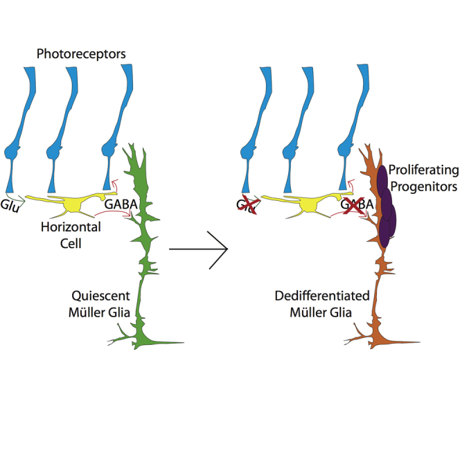
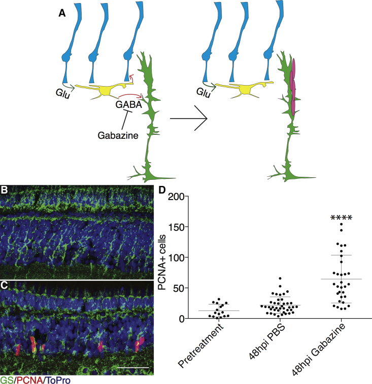
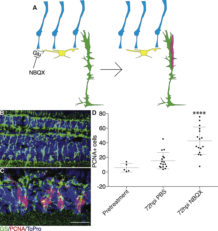
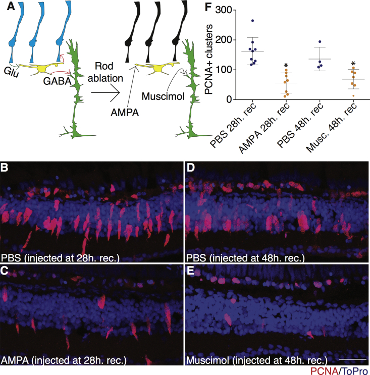
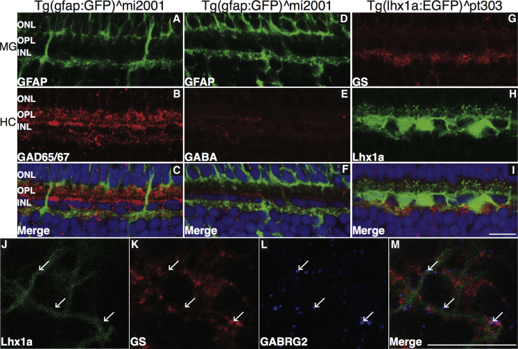
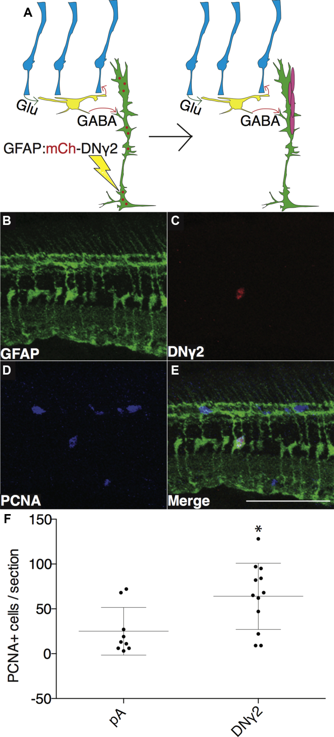
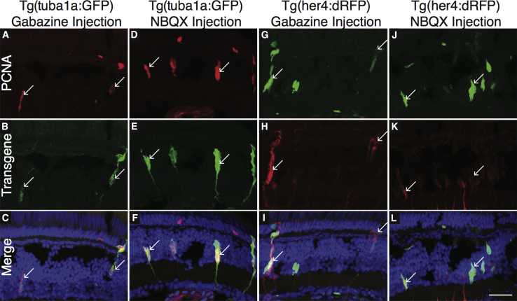
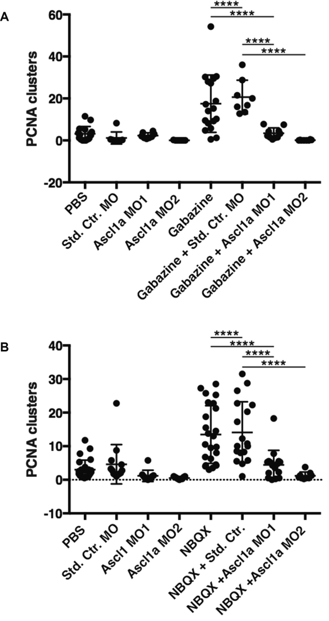
Current efforts to repair damaged or diseased mammalian retinas are inefficient and largely incapable of fully restoring vision. Conversely, the zebrafish retina is capable of spontaneous regeneration upon damage using Müller glia (MG)-derived progenitors. Understanding how zebrafish MG initiate regeneration may help develop new treatments that prompt mammalian retinas to regenerate. We show that inhibition of γ-aminobutyric acid (GABA) signaling facilitates initiation of MG proliferation. GABA levels decrease following damage, and MG are positioned to detect decreased ambient levels and undergo dedifferentiation. Using pharmacological and genetic approaches, we demonstrate that GABAA receptor inhibition stimulates regeneration in undamaged retinas while activation inhibits regeneration in damaged retinas.

Keywords: GABA; Müller glia; regeneration; retina; zebrafish.


Figures

None
Graphical abstract
Figure 1
Figure 1
Gabazine Injections Cause Time-Dependent Spontaneous Proliferation in Undamaged Retinas (A) Model illustrating effects of gabazine injection on MG proliferation. (B and C) Wild-type eyes were injected with PBS (B) or 12.5 nmol gabazine (C) into one eye. Fish recovered for 48 hr after gabazine injections before proliferation was measured. Representative images are small portions of entire retina. Scale bar, 100 μm. (D) Proliferating cells were counted across whole sections by PCNA staining for pretreatment (n = 16 eyes analyzed), PBS injection (n = 40), and gabazine injection (n = 31). hpi, hours post injection. One-way ANOVA was used; error bars denote SD; ∗∗∗∗p < 0.0001.
Figure 2
Figure 2
NBQX Injections Cause Time-Dependent Spontaneous Proliferation in Undamaged Retinas (A) Model illustrating effects of NBQX injections on MG proliferation. (B and C) Wild-type eyes were injected with PBS (B) or 25 nmol NBQX (C) into one eye. Fish recovered for 72 hr after NBQX injections (B and C) before proliferation was measured. Representative images are small portions of entire retina. Scale bar, 100 μm. (D) Proliferating cells were counted across whole sections by PCNA staining for pretreatment (n = 5 eyes analyzed), PBS injection (n = 19), and NBQX injection (n = 16). hpi, hours post injection. One-way ANOVA was used; error bars denote SD; ∗∗∗∗p < 0.0001.
Figure 3
Figure 3
AMPA and Muscimol Injections Suppress Regeneration in Damaged Retinas (A) Model illustrating effects of muscimol and AMPA injections on MG proliferation. (B–E) Tg(zop:nfsb-EGFP)nt19 fish were treated with 10 mM metronidazole for 24 hr, then allowed to recover. Fish were then anesthetized and injected with either AMPA at 28-hr recovery (C) or muscimol at 48-hr recovery (E). PBS controls were also anesthetized and injected at 28-hr (B) or 48-hr recovery (D). All injected eyes were removed at 52-hr recovery. Proliferation was assessed by PCNA staining. Representative images are small portions of the entire retina. Scale bar, 100 μm. (F) Clusters of proliferating cells were measured across entire sections for PBS at 28 hr recovery (n = 9 eyes analyzed), AMPA at 28 hr recovery (n = 8), PBS at 48 hr recovery (n = 4), and muscimol at 48 hr recovery (n = 7). Two-tailed Student’s t test was used; error bars denote SD; p < 0.05.
Figure 4
Figure 4
Close Association of MG and HC Processes in the INL (A–I) Tg(gfap:GFP)mi2001 and Tg(lhx1a:EGFP)pt303 retina sections were stained for GAD65/67 (A–C), GABA (D–F), or GS (G–I). Scale bar, 100 μm. (J–M) Co-localization of MG and HC markers was observed in the INL. Tg(lhx1a:EGFP)pt303 retinas were removed, stained for GS and GABRG2, and the area of co-localization imaged in flat mount. Arrows indicate GABRG2 puncta. Scale bar, 100 μm. See also Figure S6.
Figure 5
Figure 5
Expression of DNγ2 in MG of Undamaged Retina Causes Increased Proliferation (A) Model illustrating effects of electroporation of DNγ2 into MG on proliferation. (B–E) A GFAP:mCh-DNγ2 construct was electroporated into one retina of undamaged Tg(gfap:GFP)mi2001 fish. GFP expression (B), mCherry expression (C), and staining for PCNA (D) all co-labeled in the same cell (E). Scale bar, 100 μm. (F) Total number of PCNA-expressing cells was measured for pA electroporation (n = 9 eyes analyzed) and DNγ2 (n = 12). Two-tailed Student’s t test was used; error bars denote SD; p < 0.05. See also Figure S7.
Figure 6
Figure 6
Injection of Gabazine or NBQX into Undamaged Eyes Causes Upregulation of Factors Associated with Regeneration Gabazine (A–C, G–I) or NBQX (D–F, J–L) was injected into one eye of Tg(tuba1a:GFP) (A–F) or Tg(her4:dRFP) (G–L) fish. Fish were allowed to recover for 72 hr, after which retinas were removed and stained for PCNA. Both GFP and dRFP expression co-labeled with PCNA. Arrows indicate co-localization of transgene and PCNA. Scale bar, 100 μm.
Figure 7
Figure 7
Knockdown of Ascl1a Inhibits Retinal Proliferation Induced by Neurotransmitter Inhibition (A) Wild-type eyes were injected with PBS (n = 16), 0.75 nmol of a standard control morpholino (Std. Ctr., n = 8), or one of two morpholinos targeting Ascl1a (Ascl1a MO1 [n = 8] and Ascl1a MO2 [n = 7]). Gabazine was also injected alone (n = 18) or in combination with morpholinos (n = 8, 10, and 8, respectively). (B) Wild-type eyes were injected with PBS (n = 26), 0.75 nmol of a standard control morpholino (Std. Ctr., n = 13), or one of two morpholinos targeting Ascl1a (Ascl1a MO1 [n = 10] and Ascl1a MO2 [n = 9]). NBQX was also injected alone (n = 24) or in combination with morpholinos (n = 18, 16, and 9, respectively). Following 1–2 hr of recovery, the injected eyes were electroporated. Seventy-two hours after injection the treated eyes were removed for immunohistochemistry and scoring. Clusters of proliferating cells were counted and averaged across four whole retinal sections per eye by PCNA staining. n Values denote the number of eyes analyzed. One-way ANOVA was used; error bars = SD; ∗∗∗∗p < 0.0001.

References

    1. Andang M., Hjerling-Leffler J., Moliner A., Lundgren T.K., Castelo-Branco G., Nanou E., Pozas E., Bryja V., Halliez S., Nishimaru H. Histone H2AX-dependent GABA(A) receptor regulation of stem cell proliferation. Nature. 2008;451:460–464. - PubMed
    1. Barber A.C., Hippert C., Duran Y., West E.L., Bainbridge J.W., Warre-Cornish K., Luhmann U.F., Lakowski J., Sowden J.C., Ali R.R. Repair of the degenerate retina by photoreceptor transplantation. Proc. Natl. Acad. Sci. USA. 2013;110:354–359. - PMC - PubMed
    1. Bernardos R.L., Barthel L.K., Meyers J.R., Raymond P.A. Late-stage neuronal progenitors in the retina are radial Muller glia that function as retinal stem cells. J. Neurosci. 2007;27:7028–7040. - PMC - PubMed
    1. Bordey A. Enigmatic GABAergic networks in adult neurogenic zones. Brain Res. Rev. 2007;53:124–134. - PubMed
    1. Braun S.M., Jessberger S. Adult neurogenesis: mechanisms and functional significance. Development. 2014;141:1983–1986. - PubMed

Publication types

Substances