. 2017 May 15:152:312-329.
doi: 10.1016/j.neuroimage.2017.03.010. Epub 2017 Mar 7.

Spinal cord grey matter segmentation challenge

Affiliations

Spinal cord grey matter segmentation challenge

Ferran Prados et al. Neuroimage. .

Abstract

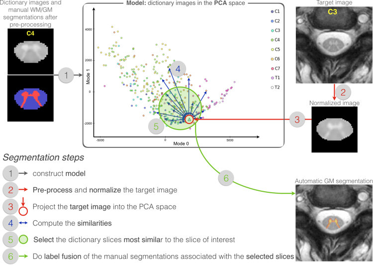
An important image processing step in spinal cord magnetic resonance imaging is the ability to reliably and accurately segment grey and white matter for tissue specific analysis. There are several semi- or fully-automated segmentation methods for cervical cord cross-sectional area measurement with an excellent performance close or equal to the manual segmentation. However, grey matter segmentation is still challenging due to small cross-sectional size and shape, and active research is being conducted by several groups around the world in this field. Therefore a grey matter spinal cord segmentation challenge was organised to test different capabilities of various methods using the same multi-centre and multi-vendor dataset acquired with distinct 3D gradient-echo sequences. This challenge aimed to characterize the state-of-the-art in the field as well as identifying new opportunities for future improvements. Six different spinal cord grey matter segmentation methods developed independently by various research groups across the world and their performance were compared to manual segmentation outcomes, the present gold-standard. All algorithms provided good overall results for detecting the grey matter butterfly, albeit with variable performance in certain quality-of-segmentation metrics. The data have been made publicly available and the challenge web site remains open to new submissions. No modifications were introduced to any of the presented methods as a result of this challenge for the purposes of this publication.

Keywords: Challenge; Evaluation metrics; Grey matter; MRI; Segmentation; Spinal cord.


Figures

Fig. 1
Fig. 1
Results of the raters for the testing dataset. Boxplot, the mean value is represented by a rhombus and dots show original obtained values per mask. Each rater's results are compared to the majority voting mask. From left to right, first row: Dice similarity coefficient (DSC), mean surface distance (MSD), Hausdorff surface distance (HSD), skeletonized median distance (SMD) and skeletonized Hausdorff distance (SHD). Second row: true positive rate (TPR), true negative rate (TNR), positive predictive value (PPV), Jaccard index (JI) and conformity coefficient (CC).
Fig. 2
Fig. 2
Dice similarity coefficient (DSC), mean surface distance (MSD), Hausdorff surface distance (HSD), skeletonized Hausdorff distance (SHD), skeletonized median distance (SMD) results of the presented methods per site using the testing dataset. Boxplot, the mean value is represented by a rhombus and dots show original obtained values per mask. MSD, HSD, SMD and SHD are in mm and represented using a logarithmic scale.
Fig. 3
Fig. 3
True positive rate (TPR), true negative rate (TNR), positive predictive value (PPV), Jaccard index (JI) and conformity coefficient (CC) results of the presented methods per site using the testing dataset. Boxplot, the mean value is represented by a rhombus and dots show original obtained values per mask.
Fig. 4
Fig. 4
Binary grey matter segmentation results for the same single slice for subject 11 of each site. From top to bottom row: input image, majority voting segmentation from the 4 raters and the segmentation methods: JCSCS, DEEPSEG, MGAC, GSBME, SCT and VBEM. Obtained 3D DSC is overlayed.
Fig. 5
Fig. 5
Schematic representation of the proposed pipeline.
Fig. 6
Fig. 6
Diagram showing the 11-layer network structure used in the present work, based on the network presented by Brosch et al. (2016). The shortcut connections between corresponding convolutional and deconvolutional layers allow for the learning of features at different scales.
Fig. 7
Fig. 7
Example of MGAC with comparisons to manual segmentations.
Fig. 8
Fig. 8
Block diagram describing the three-stage GSBME procedure for grey matter segmentation in anatomical MRI data of the spinal cord. The first step of the procedure is pre-processing, which implements whole-cord segmentation, signal intensity normalisation and image denoising. The second step is the thresholding of the sum of grey/white matter signal intensity entropies. The final stage consists of a one-class classifier for supervised outlier detection.
Fig. 9
Fig. 9
Multi-atlas based segmentation method.
Fig. 10
Fig. 10
Directed acyclic graph representing the Gaussian mixture model that the VBEM method relies on.

References

    1. Amukotuwa, S.A., Cook, M.J. (Eds.), 2015. Spinal Disease: Neoplastic, Degenerative, and Infective Spinal Cord Diseases and Spinal Cord Compression. Clinical Gate.
    1. Ashburner J., Friston K.J. Voxel-based morphometry: the methods. Neuroimage. 2000;11(6):805–821. - PubMed
    1. Ashburner J., Friston K.J. Unified segmentation. Neuroimage. 2005;26(3):839–851. - PubMed
    1. Ashburner J., Friston K.J. Nonlinear spatial normalization using basis functions. Hum. Brain Mapp. 1999;7(4):254–266. - PMC - PubMed
    1. Asman A.J., Bryan F.W., Smith S.a., Reich D.S., Landman B.a. Groupwise multi-atlas segmentation of the spinal cord's internal structure. Med. Image Anal. 2014;18(3):460–471. - PMC - PubMed

LinkOut - more resources