. 2017 Feb 24:11:88.
doi: 10.3389/fnhum.2017.00088. eCollection 2017.

Cortical Alpha Oscillations Predict Speech Intelligibility

Affiliations

Cortical Alpha Oscillations Predict Speech Intelligibility

Andrew Dimitrijevic et al. Front Hum Neurosci. .

Abstract

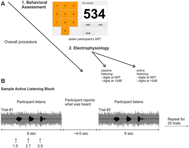
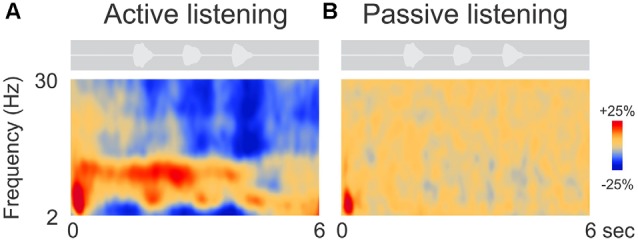
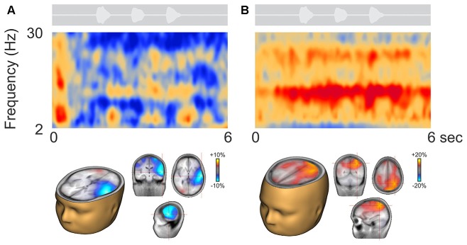
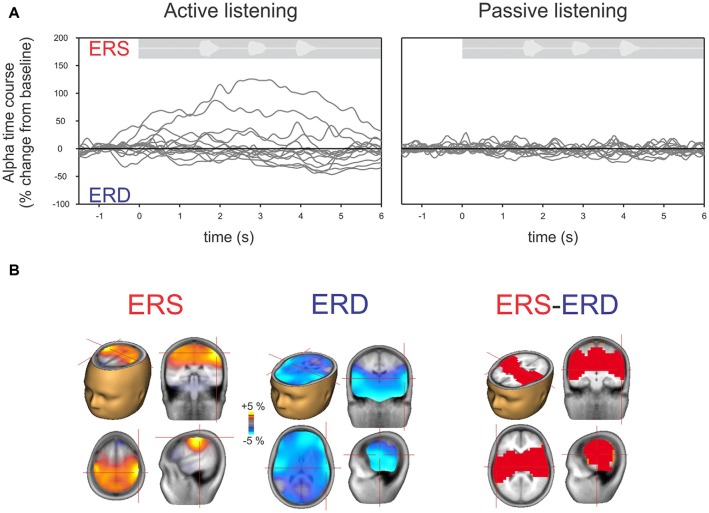
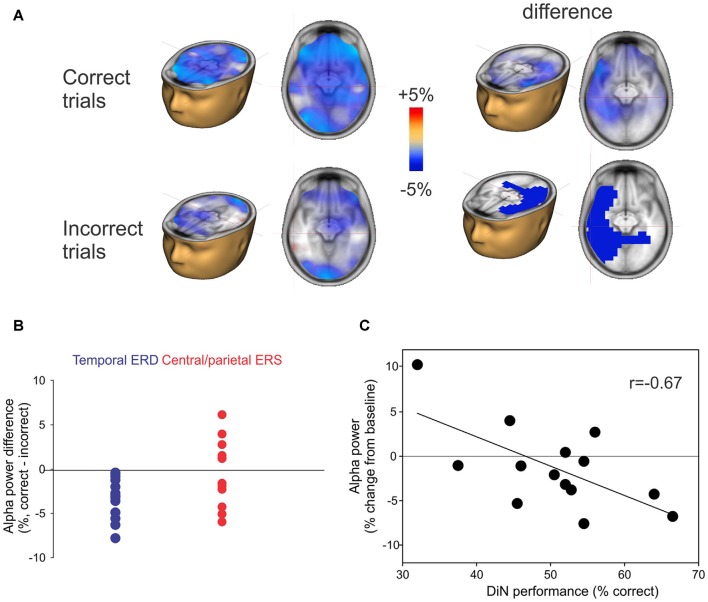
Understanding speech in noise (SiN) is a complex task involving sensory encoding and cognitive resources including working memory and attention. Previous work has shown that brain oscillations, particularly alpha rhythms (8-12 Hz) play important roles in sensory processes involving working memory and attention. However, no previous study has examined brain oscillations during performance of a continuous speech perception test. The aim of this study was to measure cortical alpha during attentive listening in a commonly used SiN task (digits-in-noise, DiN) to better understand the neural processes associated with "top-down" cognitive processing in adverse listening environments. We recruited 14 normal hearing (NH) young adults. DiN speech reception threshold (SRT) was measured in an initial behavioral experiment. EEG activity was then collected: (i) while performing the DiN near SRT; and (ii) while attending to a silent, close-caption video during presentation of identical digit stimuli that the participant was instructed to ignore. Three main results were obtained: (1) during attentive ("active") listening to the DiN, a number of distinct neural oscillations were observed (mainly alpha with some beta; 15-30 Hz). No oscillations were observed during attention to the video ("passive" listening); (2) overall, alpha event-related synchronization (ERS) of central/parietal sources were observed during active listening when data were grand averaged across all participants. In some participants, a smaller magnitude alpha event-related desynchronization (ERD), originating in temporal regions, was observed; and (3) when individual EEG trials were sorted according to correct and incorrect digit identification, the temporal alpha ERD was consistently greater on correctly identified trials. No such consistency was observed with the central/parietal alpha ERS. These data demonstrate that changes in alpha activity are specific to listening conditions. To our knowledge, this is the first report that shows almost no brain oscillatory changes during a passive task compared to an active task in any sensory modality. Temporal alpha ERD was related to correct digit identification.

Keywords: EEG; attention; brain; digits in noise; hearing; speech in noise.


Figures

Figure 1
Figure 1
(A) Overall experimental procedure. First, a behavioral assessment was performed on each participant to obtain the digits-in-noise (DiN) speech reception threshold (SRT). Afterwards, the electrophysiology was performed using the subject-specific SRT or +2 dB. (B) Two sample trials in the active listening portion of the experiment. The signal to noise ratio (SNR) was kept constant at the SRT or +2 dB. After the DiN was presented, the participant verbally reported what digits were perceived. The experimenter recorded the participant’s response then initiated the next trial. This was repeated for a total of 25 trials in each run. Eight runs were recorded in total resulting in 200 trials per condition, (for SRT, ~100 correct and ~100 correct).
Figure 2
Figure 2
Grand mean time-frequency representations of the electrophysiological data. The left (A) side shows the mean trial data from the active listening condition whereas, on the right (B), data from the passive listening are shown. The DiN stimulus is shown above each plot indicating the timing of the stimuli relative to the time-frequency plot. Time-frequency plots have a number of different illustration conventions. In this case, oscillatory changes are calculated as a percent change from baseline. Oscillatory activity is shown as increased (red; event-related synchronization, ERS) or decreased (blue; event-related desynchronization, ERD) activity relative to baseline (not shown). Note that the active condition is characterized by more oscillatory power in the alpha (8–12 Hz), beta (15–30 Hz) delta/theta (2–6 Hz) bands than in the passive listening condition. Time frequency data were averaged across all 63 electrodes.
Figure 3
Figure 3
Representative participants during active listening. Variability in the time-frequency representations were observed. Some participants, had alpha ERD that localized to temporal/auditory regions (A) while others had more ERS that localized to central or parietal regions (B). On average, the ERD was of smaller magnitude than the ERS (note the scale differences; see also Figure 4A).
Figure 4
Figure 4
(A) Time course of alpha (8–12 Hz) across individual listeners shown separately for active (left) and passive (right) listening. Each gray line represents a single subject’s 63-channel averaged alpha activity. By definition, increases above the baseline are defined as ERS and below the baseline as ERD. Note that, similar to Figure 2, hardly any deviations from the baseline are seen with passive listening compared to active listening where some show ERS and others ERD. (B) Mean source activation of alpha (8–12 Hz, over a 2–3 s window) is shown for those who had ERS and those who had ERD. The source of ERS is predominantly in the central/parietal regions whereas the ERD is predominately in the temporal/auditory regions. A statistical comparison between those with ERS vs. ERD showed that differences are mostly seen in the auditory/temporal regions vs. central/parietal regions (p < 0.05).
Figure 5
Figure 5
(A) Correct or incorrect identification of digits. Active listening trials were sorted and separately averaged depending on identification of all three digits. Significant differences in alpha sources were observed between correct and incorrect DiN trials. On the left, the alpha ERD power was greater in magnitude on the correct trials vs. incorrect trials. The difference (correct minus incorrect trials) are shown on the top right and significant (p < 0.05) clusters are shown below. (B) Individual peak difference in temporal (blue) alpha (correct minus incorrect) and central/parietal (red) alpha. The central/parietal alpha showed no consistent difference (correct vs. incorrect) whereas the temporal alpha was always greater in magnitude (more negative) on the correct than on incorrect trials. (C) A significant correlation was found between the alpha power (averaged across all trials, correct and incorrect) and DiN performance. The alpha power was computed at Talairach coordinates (−32, 4, 24), this location was found to have the peak correlation with DiN performance and alpha.

References

    1. Anderson S., Parbery-Clark A., White-Schwoch T., Kraus N. (2013). Auditory brainstem response to complex sounds predicts self-reported speech-in-noise performance. J. Speech Lang. Hear. Res. 56, 31–43. 10.1044/1092-4388(2012/12-0043) - DOI - PMC - PubMed
    1. Arnott S. R., Binns M. A., Grady C. L., Alain C. (2004). Assessing the auditory dual-pathway model in humans. Neuroimage 22, 401–408. 10.1016/j.neuroimage.2004.01.014 - DOI - PubMed
    1. Becker R., Pefkou M., Michel C. M., Hervais-Adelman A. G. (2013). Left temporal alpha-band activity reflects single word intelligibility. Front. Syst. Neurosci. 7:121. 10.3389/fnsys.2014.00047 - DOI - PMC - PubMed
    1. Bonnefond M., Jensen O. (2012). Alpha oscillations serve to protect working memory maintenance against anticipated distracters. Curr. Biol. 22, 1969–1974. 10.1016/j.cub.2012.08.029 - DOI - PubMed
    1. Colby C. L., Goldberg M. E. (1999). Space and attention in parietal cortex. Annu. Rev. Neurosci. 22, 319–349. 10.1146/annurev.neuro.22.1.319 - DOI - PubMed

LinkOut - more resources