Radiographic Evaluation of Common Pediatric Elbow Injuries
- PMID: 28286625
- PMCID: PMC5337779
- DOI: 10.4081/or.2017.7030
Radiographic Evaluation of Common Pediatric Elbow Injuries
Abstract
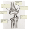
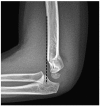
Normal variations in anatomy in the skeletally immature patient may be mistaken for fracture or injury due to the presence of secondary centers of ossification. Variations in imaging exist from patient to patient based on sex, age, and may even vary from one extremity to the other on the same patient. Despite differences in the appearance of the bony anatomy of the elbow there are certain landmarks and relationships, which can help, distinguish normal from abnormal. We review common radiographic parameters and pitfalls associated in the evaluation of pediatric elbow imaging. We also review common clinical diagnoses in this population.
Keywords: Development; Elbow; Fracture; Pediatrics; Radiographs.
Figures


References
-
- Emery KH, Zingula SN, Anton CG, et al. Pediatric elbow fractures: a new angle on an old topic. Pediatr Radiol 2016;46:61-6. - PubMed
-
- Landin LA. Fracture patterns in children. Analysis of 8,682 fractures with special reference to incidence, etiology and secular changes in a Swedish urban population 1950-1979. Acta Orthop Scand Suppl 1983;202:1-109. - PubMed
-
- Skaggs D, Pershad J. Pediatric elbow trauma. Pediatr Emerg Care. 1997;13:425-34. - PubMed
-
- Nakamura K, Hirachi K, Uchiyama S, et al. Long-term clinical and radiographic outcomes after open reduction for missed Monteggia fracture-dislocations in children. J Bone Joint Surg Am 2009;91:1394-404. - PubMed
-
- Rodgers WB, Waters PM, Hall JE. Chronic Monteggia lesions in children. Complications and results of reconstruction. J Bone Joint Surg Am 1996;78:1322-9. - PubMed
Publication types
LinkOut - more resources
Full Text Sources
Other Literature Sources

