Review
. 2017:2017:5364827.
doi: 10.1155/2017/5364827. Epub 2017 Feb 13.

Spina Bifida: Pathogenesis, Mechanisms, and Genes in Mice and Humans

Affiliations
Review

Spina Bifida: Pathogenesis, Mechanisms, and Genes in Mice and Humans

Siti W Mohd-Zin et al. Scientifica (Cairo). 2017.

Abstract

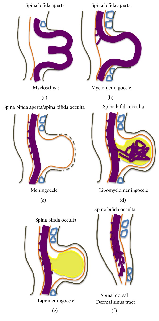
Spina bifida is among the phenotypes of the larger condition known as neural tube defects (NTDs). It is the most common central nervous system malformation compatible with life and the second leading cause of birth defects after congenital heart defects. In this review paper, we define spina bifida and discuss the phenotypes seen in humans as described by both surgeons and embryologists in order to compare and ultimately contrast it to the leading animal model, the mouse. Our understanding of spina bifida is currently limited to the observations we make in mouse models, which reflect complete or targeted knockouts of genes, which perturb the whole gene(s) without taking into account the issue of haploinsufficiency, which is most prominent in the human spina bifida condition. We thus conclude that the need to study spina bifida in all its forms, both aperta and occulta, is more indicative of the spina bifida in surviving humans and that the measure of deterioration arising from caudal neural tube defects, more commonly known as spina bifida, must be determined by the level of the lesion both in mouse and in man.


Conflict of interest statement

The authors declare that they have no competing interests.

Figures

Figure 1
Figure 1
Schematic representation of the open (aperta) and close (occulta) types of spina bifida. (a) Myeloschisis which represents the most severe form of open spina bifida. (b) Myelomeningocele which represents another typical severe form of open spina bifida (spina bifida aperta/spina bifida cystica). The typical representation is that of the spinal cord lying outside the spinal canal. (c) Meningocele that represents open or close spina bifida (the skin may or may not be present) but spinal cord does not lie outside the spinal canal. (d) Lipomyelomeningocele that represents closed spina bifida (spina bifida occulta) (covered with skin) but spinal cord is intermeshed with lipid globules (in yellow). (e) Lipomeningocele that exhibits closed spina bifida but spinal cord does not lie outside spinal canal even though lipid globules are present. (f) Spinal dorsal dermal sinus tract; spina bifida occulta with vertebral arches missing (often asymptomatic and is thought to be a mesodermal defect and a defect of secondary neurulation).
Figure 2
Figure 2
Points of closure in the mouse embryo and phenotypes of failure of closure of the various points along the neuraxis. (a) Schematic figure illustrating the multiple points of closure of the neural tube, directions of closure, and the different locations of neuropores in the developing embryo. (1), site of closure (1) which occurs at the level of somite 3 in the 6-7-somite embryo. Closure (1) is the initiation event of neurulation. Closure then progresses caudally and is completed by closure of the posterior neuropore (PNP) at the 29-30-somite stage of development; (2), second closure site at around the 10-somite stage; (3), closure (3) site which begins soon after closure (2). Arrows depict spreading of neural tube closure to neighbouring regions with completion of anterior neuropore closure soon after initiation of closure (3) and closure of the hindbrain neuropore at the 18–20-somite stage. (b) Phenotype resulting from failure of closure (1): craniorachischisis; (c) phenotype resulting from failure of closure (2): exencephaly; (d) phenotype of failure of the caudal wave of spinal closure, leading to an enlarged PNP and later development of spina bifida. (A), posterior neuropore; (B), branchial arches; (C), developing heart; (D), hindbrain; (E), midbrain; (F), forebrain; ANP: anterior neuropore; HNP: hindbrain neuropore.
Figure 3
Figure 3
Schematic representation of the formation of the mouse spinal neural tube. Process of closure of the PNP of embryos undergoing Mode 1 (a, b, d) or Mode 2 (a, c, d) neurulation. (a) Neuroepithelium thickens and converges; (b) formation of bilateral neural folds which are elevated (Mode 1); (c) apposing tips of neural folds aided by bending at the dorsolateral hinge points (DLHP) of the bilateral neural folds (Mode 2); (d) adhesion and fusion at the tips of the neural folds; (e) remodeling of the neural tube. Ne, neuroepithelium; Se, surface ectoderm; Me, mesoderm; MHP, median hinge point; DLHP, dorsolateral hinge points; POAF, point of adhesion and fusion; Nt, notochord.
Figure 4
Figure 4
Schematic figure showing progressive developmental stages of the mouse embryo and sections through the PNP at these stages. (a) Schematic of embryo at 6–10-somite stage, which has already undergone closure (1); (b) section through PNP of (a), depicting Mode 1 neurulation; (c) schematic of embryo at 12–15-somite stage; (d) section through PNP of (c) exhibiting Mode 2 neurulation; (e) schematic of embryo which has undergone closures (1), (2), and (3) with PNP being the only remaining unfused section of the neural tube; (f) section through PNP of (e) depicting Mode 3 neurulation.

Similar articles

Cited by

References

    1. Szymanski K. M., Misseri R., Whittam B., et al. Quality of life assessment in spina bifida for children (QUALAS-C): development and validation of a novel health-related quality of life instrument. Urology. 2016;87:178–184. doi: 10.1016/j.urology.2015.09.027. - DOI - PubMed
    1. Fischer N., Church P., Lyons J., Mcpherson A. C. A qualitative exploration of the experiences of children with spina bifida and their parents around incontinence and social participation. Child: Care, Health and Development. 2015;41(6):954–962. doi: 10.1111/cch.12257. - DOI - PubMed
    1. Copp A. J., Adzick N. S., Chitty L. S., Fletcher J. M., Holmbeck G. N., Shaw G. M. Spina bifida. Nature Reviews Disease Primers. 2015;1, article 15007 doi: 10.1038/nrdp.2015.7. - DOI - PMC - PubMed
    1. Harris M. J., Juriloff D. M. An update to the list of mouse mutants with neural tube closure defects and advances toward a complete genetic perspective of neural tube closure. Birth Defects Research Part A: Clinical and Molecular Teratology. 2010;88(8):653–669. doi: 10.1002/bdra.20676. - DOI - PubMed
    1. Juriloff D. M., Harris M. J. A consideration of the evidence that genetic defects in planar cell polarity contribute to the etiology of human neural tube defects. Birth Defects Research A—Clinical and Molecular Teratology. 2012;94(10):824–840. doi: 10.1002/bdra.23079. - DOI - PubMed

LinkOut - more resources