. 2017 Feb;2(1):39-53.
doi: 10.1016/j.jacbts.2016.10.006.

An Oral Selective Alpha-1A Adrenergic Receptor Agonist Prevents Doxorubicin Cardiotoxicity

Affiliations

An Oral Selective Alpha-1A Adrenergic Receptor Agonist Prevents Doxorubicin Cardiotoxicity

JuYoun Beak et al. JACC Basic Transl Sci. 2017 Feb.

Abstract

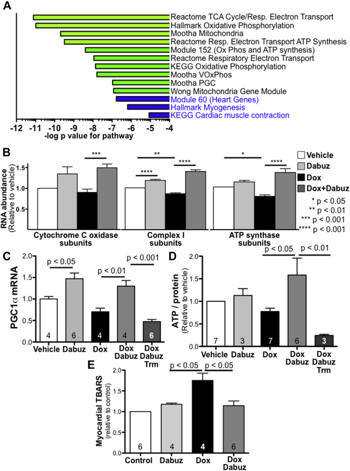
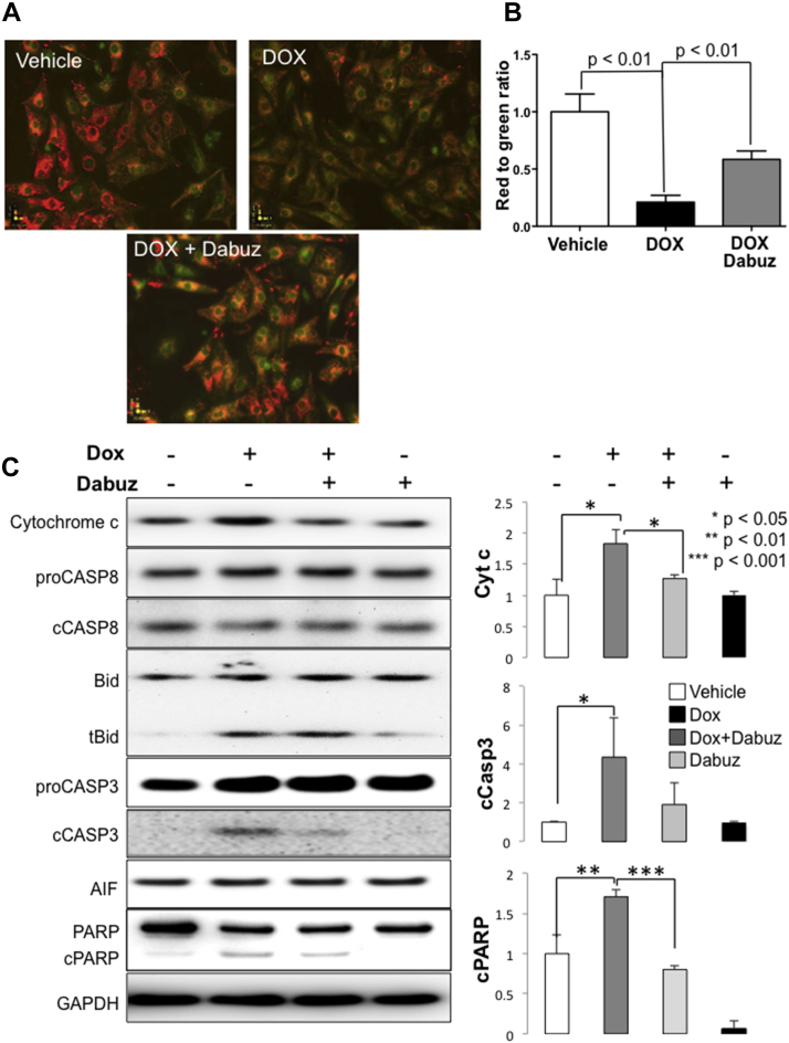
α1A-ARs play adaptive and protective roles in the heart. Dabuzalgron is an oral selective α1A-AR agonist that was well tolerated in multiple clinical trials of treatment for urinary incontinence, but has never been used to treat heart disease in humans or animal models. In this study, we administered dabuzalgron to mice treated with DOX, a widely used chemotherapeutic agent with dose-limiting cardiotoxicity that can lead to HF. Dabuzalgron protected against DOX-induced cardiotoxicity, likely by preserving mitochondrial function. These results suggest that activating cardiac α1A-ARs with dabuzalgron, a well-tolerated oral agent, might represent a novel approach to treating HF.

Keywords: adrenergic; alpha; anthracyclines; cardioprotection; catecholamines; heart failure; receptors.


Figures

None
Graphical abstract
Figure 1
Figure 1
Dabuzalgron Does Not Affect BP or Cause Myocardial Hypertrophy in Uninjured WT Mice (A) Blood pressure (BP) and heart rate (HR) were measured noninvasively in male mice for 10 days. All daily values represent the average of at least 20 cuff inflations. Mice were trained on the apparatus for the first 5 days, during which no drug was administered. On Days 6 to 10, mice were gavaged with dabuzalgron (100 ng/kg to 100 μg/kg) or water twice daily. (B) Male mice were treated with dabuzalgron 10 μg/kg by gavage twice daily for 7 days. Heart weight (HW) (in mg) was indexed to tibia length (TL) (in mm). (C) Quantitative reverse transcription polymerase chain reaction (qRT-PCR) was performed using heart tissue snap frozen at the time of sacrifice. ANP = atrial natriuretic peptide; d = day; Dabuz = dabuzalgron; MHCβ = myosin heavy chain beta; NS = nonsignificant; skAct = skeletal actin; WT = wild-type.
Figure 2
Figure 2
Dabuzalgron Protects Mice Against DOX Cardiotoxicity by Activating the α1A-AR (A) WT mice and knockout mice lacking the α1A-AR (AKO) underwent baseline awake echocardiography, received either doxorubicin (DOX) 20 mg/kg or vehicle control (VC) by intraperitoneal (IP) injection, then 7 days of treatment with either dabuzalgron 10 μg/kg or water by gavage twice daily. On Day 7, the mice underwent awake echocardiography before sacrifice. All analyses included only mice that survived to Day 7. (B) Fractional shortening, a measure of contractile function, with representative M-mode echocardiogram images. Results for mice that survived to Day 7 were compared in indicated groups using the Student t test, assuming normal distribution of values. (C) Day 7 heart sections stained with Masson Trichrome. Fibrosis (weighted average collagen content) was quantified using Aperio ImageScope software. Results were compared across treatment conditions by analysis of variance. Abbreviations as in Figure 1.
Figure 3
Figure 3
Dabuzalgron Augments Mitochondrial Transcript Expression and Function in Hearts From Mice Treated With DOX Male mice were treated with either DOX 20 mg/kg or vehicle by IP injection followed by 7 days gavage with either dabuzalgron 10 μg/kg twice daily, water, or trametinib (Trm) (1 mg/kg daily). Heart tissue was collected and immediately flash frozen on Day 7. (A) RNAseq was performed using RNA from the hearts of 3 mice per group (PBS + water; PBS + dabuzalgron; DOX + water; DOX + dabuzalgron). Gene set analysis was performed on DESeq2-derived statistics across these four categories. The results were highly enriched in gene sets involved in mitochondrial processes, a selection of which is shown here. (B) RNA abundance for all sequenced cytochrome C oxidase subunits (25 genes), mitochondrial complex I subunits (42 genes), and ATP synthase subunits (17 genes) was normalized by individual gene to vehicle treatment, then aggregated by treatment group. (C) Quantitative reverse transcription polymerase chain reaction for peroxisome proliferator-activated receptor gamma coactivator 1-alpha (PGC1α) was performed on mouse heart tissue (n in individual bars) (D) ATP content was measured in freshly harvested mouse heart tissue (total n in individual bars), then quantified relative to protein content. Results are presented relative to vehicle treatment for 4 independent experiments. (E) Thiobarbituric acid reactive substances (TBARS) were assayed in mouse myocardium. Abbreviations as in Figures 1 and 2.
Figure 4
Figure 4
Dabuzalgron Activates ERK, A Canonical Signaling Partner of the α1A-AR (A) NRVMs were pre-treated with the β-AR antagonist propranolol then exposed for 15 minutes to dabuzalgron. The non-selective α1-AR agonist norepinephrine (NE) was a positive control. Lysates were blotted for total and phospho-ERK (pERK). (B) The EC50 was calculated from 4 concentrations of Dabuzalgron across 5 separate experiments. (C) Summary of pERK/ERK for experiments using 5 different NRVM isolations. The average pERK/ERK ratio (≥2 individual wells) for each experiment was normalized to the pERK/ERK ratio for vehicle-treated NRVMs. (D and E) Mice were treated with DOX, DOX and dabuzalgron, Trm, or DOX, dabuzalgron and Trm for 7 days. Heart lysates were blotted for pERK and ERK. Results were compared using 1-way analysis of variance with Tukey post-test. (F) Mice underwent conscious echocardiography after 7 days of treatment with Trm, DOX and Trm, or DOX, Trm, and dabuzalgron. EC50 = half-maximal effective concentration; NRVM = neonatal rat ventricular myocyte; other abbreviations as in Figures 1, 2, and 3.
Figure 5
Figure 5
In NRVMs, Dabuzalgron Protects Against Apoptotic and Necrotic Cell Death Due to DOX NRVMs were treated for 4 h with doxorubicin 2μM in the presence and absence of dabuzalgron 10 μmol/l. Apoptosis was detected using fluorescein isothiocyanate–labeled Annexin V, cell death was detected using propidium iodide (PI), and nuclei were labeled with Hoechst. (A) Representative epifluorescence microscopy for each treatment condition. (B) Fluorescent intensity was analyzed using ImageJ 1.41 software (NIH, Bethesda, Maryland) for 3 independent experiments, using at least 2 wells per condition for each experiment. An average of 352 nuclei were counted in an average of 6 microscopic fields per experiment. Abbreviations as in Figures 1, 2, 3, and 4.
Figure 6
Figure 6
In NRVMs, Dabuzalgron Regulates Activators of Apoptosis and Protects Mitochondrial Membrane Potential After Treatment With DOX NRVMs were treated for 4 h with doxorubicin 2 μmol/l in the presence and absence of dabuzalgron 10 μmol/l. (A and B) Mitochondrial membrane potential was assessed using JC-1, and fluorescent intensity was quantified using a plate reader. Red indicates intact mitochondrial membrane potential; green indicates compromised mitochondrial membrane potential. Representative images (A) and summary findings (B) are presented. (C) NRVM lysates were blotted for selected regulators of apoptosis and mitochondrial cell death effectors. Representative Western blots and summary findings from 3 independent experiments with at least 2 wells per condition in each experiment are shown. Abbreviations as in Figures 1, 2, 3, and 4.

References

    1. O’Connell T.D., Jensen B.C., Baker A.J., Simpson P.C. Cardiac alpha1-adrenergic receptors: novel aspects of expression, signaling mechanisms, physiologic function, and clinical importance. Pharmacol Rev. 2014;66:308–333. - PMC - PubMed
    1. Turnbull L., McCloskey D.T., O'Connell T.D., Simpson P.C., Baker A.J. Alpha 1-adrenergic receptor responses in alpha 1AB-AR knockout mouse hearts suggest the presence of alpha 1D-AR. Am J Physiol Heart Circ Physiol. 2003;284:H1104–H1109. - PubMed
    1. Jensen B., Swigart P., Laden M.-E., DeMarco T., Hoopes C., Simpson P. The alpha-1D is the predominant alpha-1-adrenergic receptor in human epicardial coronary arteries. J Am Coll Cardiol. 2009;54:1137–1145. - PMC - PubMed
    1. Lin F., Owens W.A., Chen S. Targeted alpha(1A)-adrenergic receptor overexpression induces enhanced cardiac contractility but not hypertrophy. Circ Res. 2001;89:343–350. - PubMed
    1. Rorabaugh B.R., Ross S.A., Gaivin R.J. alpha1A- but not alpha1B-adrenergic receptors precondition the ischemic heart by a staurosporine-sensitive, chelerythrine-insensitive mechanism. Cardiovasc Res. 2005;65:436–445. - PubMed