Review
. 2018 Apr;33(4):553-571.
doi: 10.1007/s00467-017-3629-0. Epub 2017 Mar 13.

The molecular biology of pelvi-ureteric junction obstruction

Affiliations
Review

The molecular biology of pelvi-ureteric junction obstruction

Laura Jackson et al. Pediatr Nephrol. 2018 Apr.

Abstract

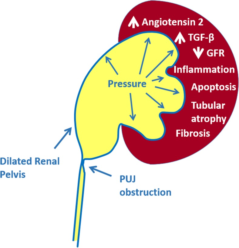
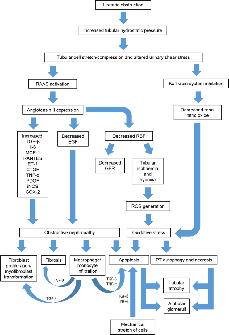
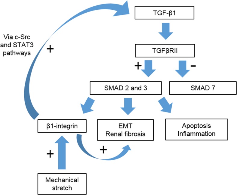
Over recent years routine ultrasound scanning has identified increasing numbers of neonates as having hydronephrosis and pelvi-ureteric junction obstruction (PUJO). This patient group presents a diagnostic and management challenge for paediatric nephrologists and urologists. In this review we consider the known molecular mechanisms underpinning PUJO and review the potential of utilising this information to develop novel therapeutics and diagnostic biomarkers to improve the care of children with this disorder.

Keywords: Aetiology; Biomarker; Hydronephrosis; Molecular biology; Pelvi-ureteric junction obstruction.


Conflict of interest statement

Funding

Laura Jackson is funded by a joint Royal College of Surgeons of England/British Association of Paediatric Surgeons Fellowship (Awarded 2014). Richard Coward is funded by the Medical Research Council as a Senior Clinical Fellow MR/K010492/1.

Conflict of interest

The authors declare that they have no conflict of interest.

Figures

Fig. 1
Fig. 1
Diagrammatic representation of the pelvi-ureteric junction (PUJ). The gradual transition from the renal pelvis to the proximal ureter is illustrated as well as the increased mucosal folds and smooth muscle thickening in this region
Fig. 2
Fig. 2
Embryological signalling pathways of the PUJ. The ureteric bud arises from the mesonephric duct and initially consists of only a simple epithelial layer extending into loose mesenchyme. Epithelial cell proliferation and differentiation to form transitional epithelium leads to luminal obliteration, which at the end of the embryonic period is corrected by physiologic recanalisation of the ureter. Epithelial paracrine and mesenchymal autocrine signalling stimulates the proliferation and differentiation of the mesenchyme into smooth muscle cells (SMC) which aggregate and orientate so as to encircle the epithelial tube. Specifically, the urothelium secretes SHH which activates the PTCH1 receptor on adjacent mesenchyme, thereby stimulating mesenchymal proliferation. Mesenchymal cells (MC) express TBX18, a T-box transcription factor, which enables the correct localisation and aggregation of the former around the urothelium. The mesenchymal cells also express BMP4 which acts in an autocrine manner to upregulate TSHZ3 and MYODC. MYODC enables differentiation of SMC by increasing the transcription of genes encoding smooth muscle contractile proteins. DLGH1, expressed by the urothelium and SMC, is responsible for the correct orientation of SMC around the urothelial tube. In postnatal mice (equivalent to second trimester of gestation in humans), increased urine production matches the development of the renal pelvis and is accompanied by a second phase of muscle differentiation that particularly affects the renal pelvis and proximal ureter, regulated by calcineurin and angiotensin II signalling. The timeline refers to days of gestation (E embryonic day) in mouse models. MD Mesonephric duct, UB ureteric bud, MC mesenchymal cells. See Table 1 for description of factors active in the pathways involved in ureteric development
Fig. 3
Fig. 3
Pathologic features of intrinsic PUJO. Reduced luminal mucosal folds, excess collagen deposition, depletion of nerves within the muscular layer, abnormal muscle fibre arrangement, inflammatory infiltrate and both muscle hypertrophy/hyperplasia and muscle atrophy/hypoplasia are seen at the PUJ in human PUJO
Fig. 4
Fig. 4
Pathologic features of rodent models of unilateral ureteric obstruction (UUO). Timeline of the development of renal pathogenic features in neonatal and adult models of UUO. CUUO Complete UUO, PUUO partial UUO
Fig. 5
Fig. 5
Major mechanisms of renal injury in PUJO. GFR glomerular filtration rate, TGF transforming growth factor
Fig. 6
Fig. 6
Major pathways involved in the development of obstructive nephropathy derived from animal and human studies. ET-1 Endothelin 1, iNOS inducible nitric oxide synthase, PT proximal tubule, RAAS renin–angiotensin–aldosterone system, RANTES regulated on activation normal T-cell expressed and secreted, RBF renal blood flow, ROS reactive oxygen species. For other abbreviations, see footnotes to Tables 3 and 4
Fig. 7
Fig. 7
Transforming growth factor β1 (TGF-β1) signalling via the SMAD-dependent pathway. Unilateral ureteric obstruction induces increased TGF-β1 and TGF-β receptor II (TGFβRII) expression, upregulating SMAD 2 and 3 and downregulating SMAD 7 (inhibitory for SMAD 2 and 3). β1-integrin is upregulated by both SMAD signalling and mechanical stretch and contributes to a positive feedback loop regulating TGF-β1 expression via the c-SRC and STAT-3 pathways. EMT Epithelial mesenchymal transformation

References

    1. Dudley JA, Haworth JM, McGraw ME, Frank JD, Tizard EJ. Clinical relevance and implications of antenatal hydronephrosis. Arch Dis Child Fetal Neonatal Ed. 1997;76:F31–34. doi: 10.1136/fn.76.1.F31. - DOI - PMC - PubMed
    1. Brussels Free University Perinatal Nephrology Study Group. Ismaili K, Avni FE, Wissing KM, Hall M. Long-term clinical outcome of infants with mild and moderate fetal pyelectasis: validation of neonatal ultrasound as a screening tool to detect significant nephrouropathies. J Pediatr. 2004;144:759–765. - PubMed
    1. Jaswon MS, Dibble L, Puri S, Davis J, Young J, Dave R, Morgan H. Prospective study of outcome in antenatally diagnosed renal pelvis dilatation. Arch Dis Child Fetal Neonatal Ed. 1999;80:F135–138. doi: 10.1136/fn.80.2.F135. - DOI - PMC - PubMed
    1. Woodward M, Frank D. Postnatal management of antenatal hydronephrosis. BJU Int. 2002;89:149–156. doi: 10.1046/j.1464-4096.2001.woodward.2578.x. - DOI - PubMed
    1. Morris RK, Kilby MD. Congenital urinary tract obstruction. Best Pract Res Clin Obstet Gynaecol. 2008;22:97–122. doi: 10.1016/j.bpobgyn.2007.08.007. - DOI - PubMed

Publication types

MeSH terms

Supplementary concepts

LinkOut - more resources