. 2017 Mar 13:8:14768.
doi: 10.1038/ncomms14768.

Brigatinib combined with anti-EGFR antibody overcomes osimertinib resistance in EGFR-mutated non-small-cell lung cancer

Affiliations

Brigatinib combined with anti-EGFR antibody overcomes osimertinib resistance in EGFR-mutated non-small-cell lung cancer

Ken Uchibori et al. Nat Commun. .

Abstract

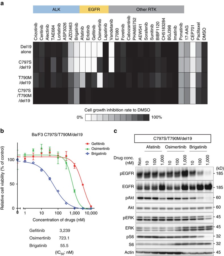
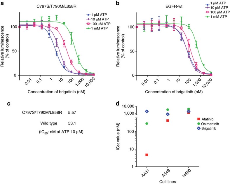
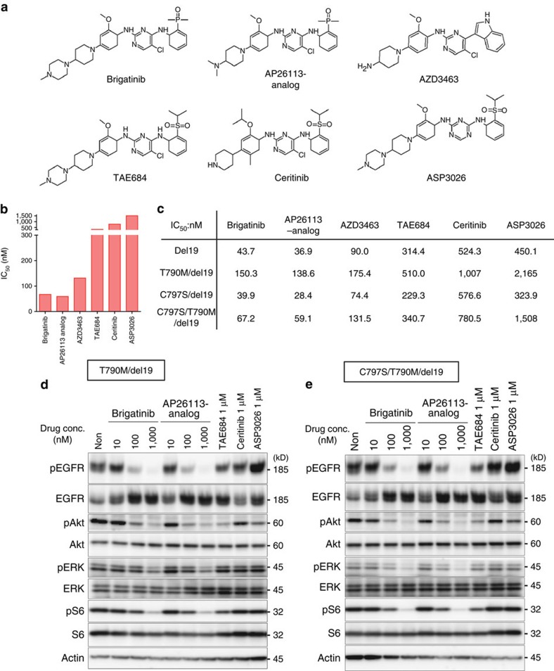
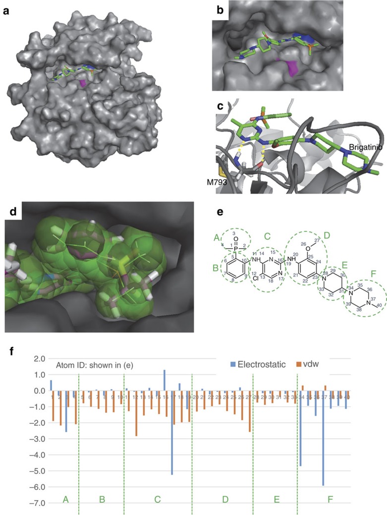
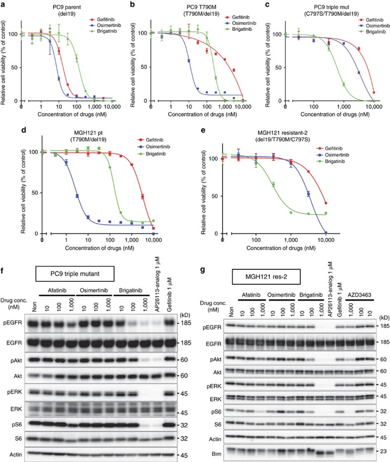
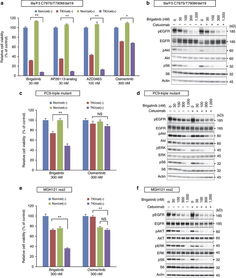
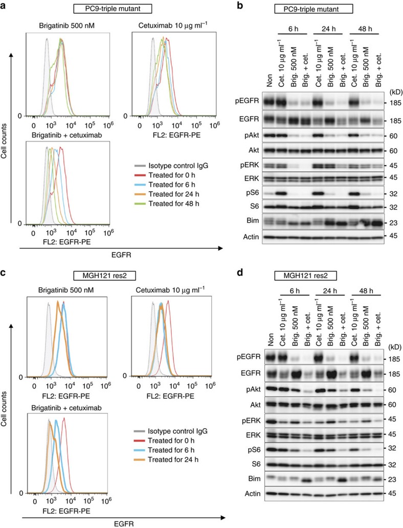
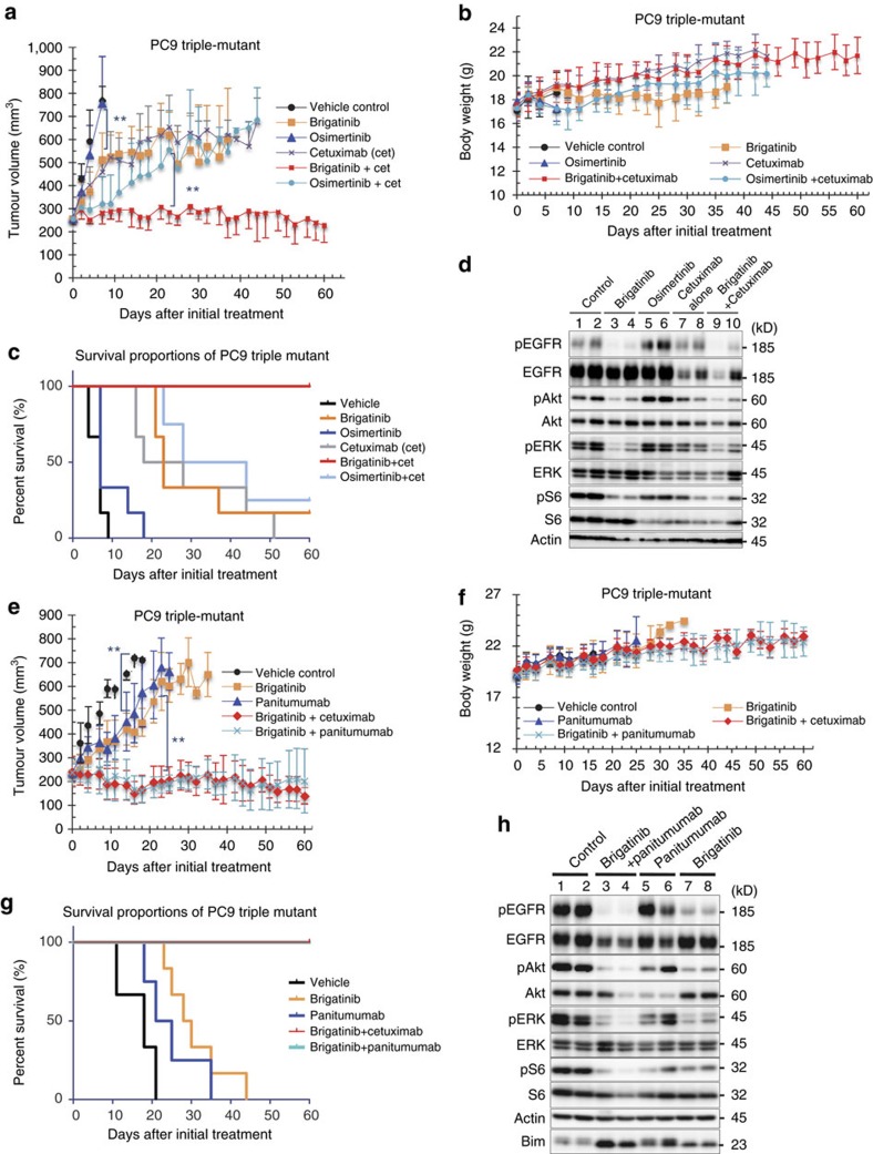
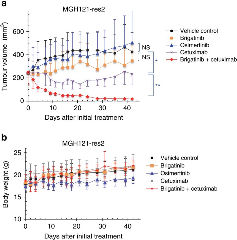
Osimertinib has been demonstrated to overcome the epidermal growth factor receptor (EGFR)-T790M, the most relevant acquired resistance to first-generation EGFR-tyrosine kinase inhibitors (EGFR-TKIs). However, the C797S mutation, which impairs the covalent binding between the cysteine residue at position 797 of EGFR and osimertinib, induces resistance to osimertinib. Currently, there are no effective therapeutic strategies to overcome the C797S/T790M/activating-mutation (triple-mutation)-mediated EGFR-TKI resistance. In the present study, we identify brigatinib to be effective against triple-mutation-harbouring cells in vitro and in vivo. Our original computational simulation demonstrates that brigatinib fits into the ATP-binding pocket of triple-mutant EGFR. The structure-activity relationship analysis reveals the key component in brigatinib to inhibit the triple-mutant EGFR. The efficacy of brigatinib is enhanced markedly by combination with anti-EGFR antibody because of the decrease of surface and total EGFR expression. Thus, the combination therapy of brigatinib with anti-EGFR antibody is a powerful candidate to overcome triple-mutant EGFR.


Conflict of interest statement

The authors declare no competing financial interests.

Figures

Figure 1
Figure 1. Identification of brigatinib as an EGFR-C797S/T790M/activating-mutation (triple-mutant EGFR) inhibitor.
(a) The results of screening the growth-inhibitory activity of 30 drugs in Ba/F3 cells expressing four types of EGFR-del19 with or without T790M or C797S mutations are shown in a heat map. Ba/F3 cells expressing each EGFR mutant were treated with 100 nM of the indicated inhibitors. After 72 h of drug treatment, the cell viability was measured using the CellTiter-Glo assay. Relative cell viability was calculated from each value divided by the DMSO control. Among the inhibitors, only brigatinib and ponatinib were sufficiently efficacious against the triple-mutant EGFR. AZD3463 acted as a weak inhibitor to the triple mutation. (b) Growth inhibition assessed by the CellTiter-Glo assay of EGFR-C797S/T790M/del19 (triple-del19)-mutated Ba/F3 cells treated with gefitinib, osimertinib and brigatinib.; N=3. Results are expressed as mean±s.d. IC50 values were calculated using growth inhibition assay. (c) Phosphorylation of EGFR and downstream signals were significantly inhibited by brigatinib in Ba/F3 cells expressing triple-del19 even though afatinib and osimertinib did not suppress at all the EGFR signalling of triple-del19.
Figure 2
Figure 2. Brigatinib inhibited EGFR through ATP competition and was less potent to wild-type EGFR or non-EGFR-mutated cells.
(ac) The evaluation of the inhibitory activity of brigatinib in the in vitro kinase assay using the ADP-Glo assay kit showed a dose-dependent decrease in EGFR activity with brigatinib according to the increase of ATP concentration in either (a) EGFR-C797S/T790M/L858R or (b) wild type; N=3. Results are expressed as mean±s.d. (c) IC50 value calculated at an ATP concentration of 10 μM suggested the better affinity of brigatinib to EGFR-C797S/T790M/L858R than to wild-type EGFR. (d) IC50 values calculated from the cell viability assay of non-EGFR-mutated cell lines, A431, A549 and H460, assessed using CellTiter-Glo assay kit are shown with a dot plot.
Figure 3
Figure 3. Efficacy of brigatinib and similarly structured drugs in the EGFR-mutated Ba/F3 cells and their chemical structures.
(a) Chemical structures of six ALK–TKIs were very similar. (b,c) IC50 values in Ba/F3 cells expressing four mutation types of EGFR-del19 were obtained by treatment with brigatinib, AP26113-analog, AZD3463, TAE684, ceritinib and ASP3026 for 72 h. Those of C797S/T790M/del19 were shown by bar graph (b) and those of all mutation types were demonstrated by a table (c). The CellTiter-Glo assay was used to measure cell viability. (d,e) Ba/F3 cells expressing T790M/del19 (d) or C797S/T790M/del19 (e) were treated with the indicated concentrations of brigatinib, AP26113 analog, TAE684, ceritinib or ASP3026 for 6 h. Phosphorylation of EGFR and its downstream signals were evaluated by western blotting with the indicated antibodies.
Figure 4
Figure 4. Structure model of EGFR–brigatinib interactions.
(a,b) The brigatinib-binding mode for the EGFR-C797S/T790M/L858R mutant (EGFR-triple-L858R). The mean structure of the EGFR–brigatinib complex, generated by molecular dynamic simulations for 10 docking poses, is shown. EGFR was depicted by a surface model (T790M, blue; C797S, purple; others, grey), and brigatinib was depicted by sticks (C, green; N, blue; O, red; P, orange; and H, hydrogen). In the structure model, brigatinib fits into the ATP-binding pocket without a sterical crush to T790M and C797S demonstrated by overview (a) and zoom-in of ATP-binding pocket (b). (c) Hydrogen bonds between the triple-mutant EGFR and brigatinib. The protein backbone and M793 of EGFR-triple-L858R were depicted by a grey backbone tube and sticks (C, grey; N, blue; O, red and H, hydrogen), respectively. Hydrogen bonds were shown by dashed yellow lines. (d) Comparison of the inhibitor-binding mode between the EGFR–brigatinib and ALK–TAE684 complexes. TAE684 was depicted by thick sticks (C, magenta; N, blue; O, red; S, yellow; and H, hydrogen) after the crystal structure of EML4-ALK in complex with TAE684 (PDB-ID: 2XB7) was superimposed to the modelling structure of EGFR (a grey surface model) in complex with brigatinib (a space-filling model with thin sticks). (e) Substructure and atom IDs in the energy plot were assigned to the chemical structure of brigatinib. (f) The mean interaction energy between the EGFR-triple-L858R and a brigatinib atom was calculated using molecular dynamic trajectories for 10 docking poses. Negative and positive values indicate favourable and repulsive interactions, respectively.
Figure 5
Figure 5. Inhibition of cell growth and downstream signal pathway in lung cancer cell lines by brigatinib.
(ae) PC9 (del19) (a), PC9-T790M (T790M/del19) (b), PC9-triple mutant (C797S/T790M/del19) (c), MGH121 parent (T790M/del19) (d) and MGH121 resistant-2 (C797S/T790M/del19) (e) cells were treated with serially diluted gefitinib, osimertinib and brigatinib for 72 h. Cell viability was measured using the CellTiter-Glo assay.; N=3. Results are expressed as mean±s.d. (f) Western blotting of PC9 triple mutant (C797S/T790M/del19) cells indicated that brigatinib and AP26113 analog, but not afatinib or osimertinib, suppressed phosphorylation of EGFR and its downstream signalling. (g) Similar results were obtained in MGH121 resistant-2.
Figure 6
Figure 6. Brigatinib combined with cetuximab synergistically suppressed the growth of EGFR-C797S/T790M/del19-expressing cells in vitro.
(a) The cell growth inhibition of Ba/F3 cells expressing EGFR-C797S/T790M/del19 (EGFR-triple-del19) treated with brigatinib, AP26113-analog, AZD3463 and osimertinib at indicated concentrations combined with or without cetuximab (10 μg ml−1) for 72 h assessed by CellTiter-Glo assay. (b) Inhibition of EGFR signal pathway in BaF3 EGFR-triple-del19 cells treated with brigatinib+cetuximab (10 μg ml−1) for 6 h was evaluated using western blotting. (c,d) The cell growth inhibition of PC9 triple-mutant cells (c) and MGH121-res2 cells (d) treated with brigatinib and osimertinib at indicated concentrations combined with or without cetuximab (10 μg ml−1) for 72 h assessed by CellTiter-Glo assay. (e,f) Inhibition of EGFR signal pathway in PC9 triple-mutant cells (e) and MGH121-res2 cells (f) treated with brigatinib+cetuximab (10 μg ml−1) for 6 h was evaluated using western blotting.; Results in a,c,e are expressed as mean±s.d. (N=3). The significance of difference between indicated groups are calculated by Student's t-test (NS; not significant, *P<0.05, **P<0.01).
Figure 7
Figure 7. Brigatinib combined with cetuximab enhanced internalization and reduced EGFR expression.
(a) FACS analysis using a PE-conjugated EGFR antibody of PC9 triple-mutant cells treated with brigatinib, cetuximab, brigatinib+cetuximab for 0, 6, 24 and 48 h demonstrated a time-dependent marked decrease in surface EGFR after treatment with brigatinib+cetuximab over a period of up to 48 h, and a moderate decrease with cetuximab alone. (b) Western blotting assessment of the cells corresponding to the treatments in a. (c,d) FACS analysis and western blotting performed with MGH121-res2 cells using the same method as with the PC9 triple-mutant cells in a,b.
Figure 8
Figure 8. Brigatinib combined with cetuximab or panitumumab synergistically suppressed the growth of EGFR-C797S/T790M/del19-expressing cells in vivo.
(a,b) PC9 cells expressing EGFR-C797S/T790M/del19 were subcutaneously implanted into Balb-c nu/nu mice. When the average tumour volume reached ∼200 mm3, the mice were randomized into vehicle control or treatment groups (50 mg kg−1 of osimertinib, 75 mg kg−1 of brigatinib, 1 mg per mouse of cetuximab three times a week or 75 mg kg−1 of brigatinib combined with cetuximab administered as previously described) and treated once daily by oral gavage for the indicated period. Tumour volume (V) was calculated as 0.5 × length × width2, and body weights (B.W.) of mice were measured twice weekly.; N=6. Results are expressed as mean±s.d. The significance of difference between the mean tumour volume of control and of brigatinib on day 7, between brigatinib and brigatinib+cetuximab on day 23, respectively, are calculated by Mann–Whitney U test (**P<0.01). (c) Survival periods of mice in each treatment arm were demonstrated using the Kaplan–Meier curve. (d) Phosphorylation of EGFR and its downstream signalling in two tumour samples obtained from each group were evaluated using western blotting. (e,f) In vivo experiment of PC9 triple-mutant cells following a similar protocol as in Fig. 8a–b, using panitumumab 0.5 mg per mouse two times a week administered peritoneally instead of cetuximab.; N=6. Results are expressed as mean±s.d. The significance of difference between the mean tumour volume of control and of brigatinib on day 16, between brigatinib and brigatinib+panitumumab on day 23, respectively, are calculated by Mann–Whitney U test (**P<0.01). (g) A Kaplan–Meier curve of the survival of the mice in each treatment arm. (h) Phosphorylation of EGFR and its downstream signalling in two tumour samples obtained from xenografts of PC9-triple mutant cells treated for 8 days with the indicated drugs (brigatinib: 75 mg kg−1 daily, administered orally; panitumumab: 0.5 mg per mouse two times a week, administered peritoneally) were assessed by western blotting with the indicated antibodies.
Figure 9
Figure 9. Brigatinib combined with cetuximab synergistically suppressed the growth of EGFR-C797S/T790M/del19-expressing lung cancer cells in vivo.
(a,b) MGH121-res2 expressing EGFR-C797S/T790M/del19 were subcutaneously implanted into SCID-beige mice. When the average tumour volume reached ∼200 mm3, the mice were randomized into vehicle control and treatment groups (50 mg kg−1 of osimertinib (po), 75 mg kg−1 of brigatinib (po), 1 mg per mouse of cetuximab two times a week and 75 mg kg−1 of brigatinib combined with cetuximab administered as previously described, respectively) and treated for the indicated period. Tumour volume (V) was calculated as 0.5 × length × width2, and the body weights (B.W.) of the mice were measured twice weekly. N=6. Results are expressed as mean±s.d. The significance in difference between the mean tumour volume of control and of osimertinib, brigatinib and cetuximab, between cetuximab and brigatinib+cetuximab, respectively, on day 42 are calculated by Mann–Whitney U test (NS: not significant, *P<0.05, **P<0.01).

References

    1. Kohno T. et al.. RET fusion gene: translation to personalized lung cancer therapy. Cancer Sci. 104, 1396–1400 (2013). - PMC - PubMed
    1. Giaccone G. Epidermal growth factor receptor inhibitors in the treatment of non-small-cell lung cancer. J. Clin. Oncol. 23, 3235–3242 (2005). - PubMed
    1. Maemondo M. et al.. Gefitinib or chemotherapy for non-small-cell lung cancer with mutated EGFR. N. Engl. J. Med. 362, 2380–2388 (2010). - PubMed
    1. Mok T. S. et al.. Gefitinib or carboplatin-paclitaxel in pulmonary adenocarcinoma. N. Engl. J. Med. 361, 947–957 (2009). - PubMed
    1. Mitsudomi T. et al.. Gefitinib versus cisplatin plus docetaxel in patients with non-small-cell lung cancer harbouring mutations of the epidermal growth factor receptor (WJTOG3405): an open label, randomised phase 3 trial. Lancet Oncol. 11, 121–128 (2010). - PubMed

Publication types

MeSH terms